Product Citations: 569
Powered by

Tachykinin signaling defines distinct populations of glia in the enteric nervous system.
In Neuron on 18 March 2026 by Muppirala, A. N., Mitchell, P. E., et al.
In Journal of Pain Research on 25 February 2026 by Shi, W., Wang, M., et al.
Activation of posterior ventral pallidum by bitter taste in mice.
In MicroPublication. Biology on 30 January 2026 by Tanaka, D. H. & Tanabe, T.
In Front Behav Neurosci on 15 March 2013 by Kobayashi, Y., Sano, Y., et al.
Fig.1.B
-
IHC-IF
-
Mus musculus (House mouse)
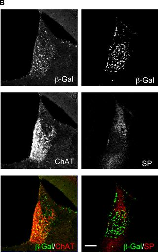
Characterization of mHb-specific Cre expression transgenic mice. Transgene combinations are schematically illustrated at the top of the figure (A,F). (A) β-galactosidase (β-Gal) activity (blue signals) in Gpr151-Cre:RNZ mouse brain sections. The left and right panels show sagittal and coronal sec...
more
Characterization of mHb-specific Cre expression transgenic mice. Transgene combinations are schematically illustrated at the top of the figure (A,F). (A) β-galactosidase (β-Gal) activity (blue signals) in Gpr151-Cre:RNZ mouse brain sections. The left and right panels show sagittal and coronal sections, respectively. Blue signals represent nuclei of cells expressing Cre protein under a Gpr151 promoter. The medial habenula (mHb) and lateral habenula (lHb) are encircled by red and blue lines, respectively. (B) Protein localization of β-Gal, choline acetyltransferase (ChAT), and substance P (SP) in the Hb of Gpr151-Cre:RNZ mice. (C) Cre-mediated recombination occurred preferentially in the ventral area of the mHb, across the entire anteroposterior axis of the habenular nuclei. Only a small population of lHb neurons in the posterior area exhibited recombination. (D) The majority of cells labeled with β-Gal protein (brown) in the lHb were characterized by Chrm2 (muscarinic acetylcholine receptor 2; M2) (blue) expression in Gpr151-Cre:RNZ mice. The mHb and lHb are outlined by red and blue lines, respectively. (E) β-Gal staining on postnatal days (P) 7, 10, 14, and 18, and 6 w. Recombination mediated by Cre protein started between P7 and P10 and progressively increased by P18. (F–H) LacZ staining of Gpr151-Cre:RGZ mice revealed β-Gal activity in the fasciculus retroflexus (fr), but not in the monoaminergic centers such as the ventral tegmental area (VTA), substantia nigra (SN), and medial raphe nucleus (MnR). (F) Blue signals represent axons originating from recombined cells. These cells project to the IPN through the fr. Representations of the fr, VTA, and SN (G), and MnR (H) are indicated by blue lines. MM, mammillary nucleus. Scale bars = 1 mm (A,G,H), 500 μm (C,E,F), 100 μm (B), and 50 μm (D).
less
Collected and cropped from Frontiers in Behavioral Neuroscience by CiteAb, provided under a CC-BY license
Image 1 of 1