Circulating monocytes are recruited in damaged tissues to generate macrophages that modulate disease progression. Colony-stimulating factor-1 (CSF-1) promotes the generation of monocyte-derived macrophages, which involves caspase activation. Here, we demonstrate that activated caspase-3 and caspase-7 are located to the vicinity of the mitochondria in CSF1-treated human monocytes. Active caspase-7 cleaves p47PHOX at aspartate 34, which promotes the formation of the NADPH (nicotinamide adenine dinucleotide phosphate) oxidase complex NOX2 and the production of cytosolic superoxide anions. Monocyte response to CSF-1 is altered in patients with a chronic granulomatous disease, which are constitutively defective in NOX2. Both caspase-7 down-regulation and radical oxygen species scavenging decrease the migration of CSF-1-induced macrophages. Inhibition or deletion of caspases prevents the development of lung fibrosis in mice exposed to bleomycin. Altogether, a non-conventional pathway that involves caspases and activates NOX2 is involved in CSF1-driven monocyte differentiation and could be therapeutically targeted to modulate macrophage polarization in damaged tissues.
Product Citations: 36
Applications
Reactivity
Research Area
Caspase Inhibition Modulates Monocyte-Derived Macrophage Polarization in Damaged Tissues.
In International Journal of Molecular Sciences on 19 February 2023 by Solier, S., Mondini, M., et al.
-
Immunology and Microbiology
In The Journal of Biological Chemistry on 5 August 2016 by Vanderperre, B., Cermakova, K., et al.
Selective transport of pyruvate across the inner mitochondrial membrane by the mitochondrial pyruvate carrier (MPC) is a fundamental step that couples cytosolic and mitochondrial metabolism. The recent molecular identification of the MPC complex has revealed two interacting subunits, MPC1 and MPC2. Although in yeast, an additional subunit, MPC3, can functionally replace MPC2, no alternative MPC subunits have been described in higher eukaryotes. Here, we report for the first time the existence of a novel MPC subunit termed MPC1-like (MPC1L), which is present uniquely in placental mammals. MPC1L shares high sequence, structural, and topological homology with MPC1. In addition, we provide several lines of evidence to show that MPC1L is functionally equivalent to MPC1: 1) when co-expressed with MPC2, it rescues pyruvate import in a MPC-deleted yeast strain; 2) in mammalian cells, it can associate with MPC2 to form a functional carrier as assessed by bioluminescence resonance energy transfer; 3) in MPC1 depleted mouse embryonic fibroblasts, MPC1L rescues the loss of pyruvate-driven respiration and stabilizes MPC2 expression; and 4) MPC1- and MPC1L-mediated pyruvate imports show similar efficiency. However, we show that MPC1L has a highly specific expression pattern and is localized almost exclusively in testis and more specifically in postmeiotic spermatids and sperm cells. This is in marked contrast to MPC1/MPC2, which are ubiquitously expressed throughout the organism. To date, the biological importance of this alternative MPC complex during spermatogenesis in placental mammals remains unknown. Nevertheless, these findings open up new avenues for investigating the structure-function relationship within the MPC complex.
© 2016 by The American Society for Biochemistry and Molecular Biology, Inc.
-
Mus musculus (House mouse)
-
Biochemistry and Molecular biology
-
Cell Biology
Drp1-dependent mitochondrial fission via MiD49/51 is essential for apoptotic cristae remodeling.
In The Journal of Cell Biology on 29 February 2016 by Otera, H., Miyata, N., et al.
Mitochondrial fission facilitates cytochrome c release from the intracristae space into the cytoplasm during intrinsic apoptosis, although how the mitochondrial fission factor Drp1 and its mitochondrial receptors Mff, MiD49, and MiD51 are involved in this reaction remains elusive. Here, we analyzed the functional division of these receptors with their knockout (KO) cell lines. In marked contrast to Mff-KO cells, MiD49/MiD51-KO and Drp1-KO cells completely resisted cristae remodeling and cytochrome c release during apoptosis. This phenotype in MiD49/51-KO cells, but not Drp1-KO cells, was completely abolished by treatments disrupting cristae structure such as OPA1 depletion. Unexpectedly, OPA1 oligomers generally thought to resist cytochrome c release by stabilizing the cristae structure were similarly disassembled in Drp1-KO and MiD49/51-KO cells, indicating that disassembly of OPA1 oligomers is not directly linked to cristae remodeling for cytochrome c release. Together, these results indicate that Drp1-dependent mitochondrial fission through MiD49/MiD51 regulates cristae remodeling during intrinsic apoptosis.
© 2016 Otera et al.
-
Cell Biology
In The Journal of Biological Chemistry on 18 December 2015 by Yue, J., Ben Messaoud, N., et al.
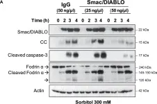
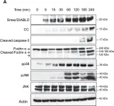
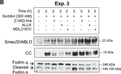
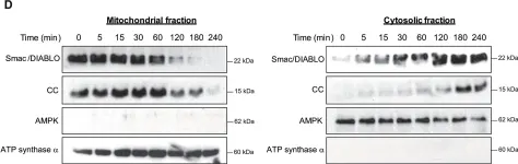
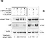
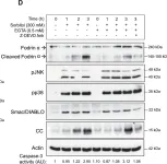
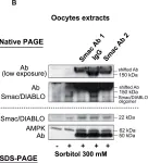
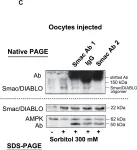
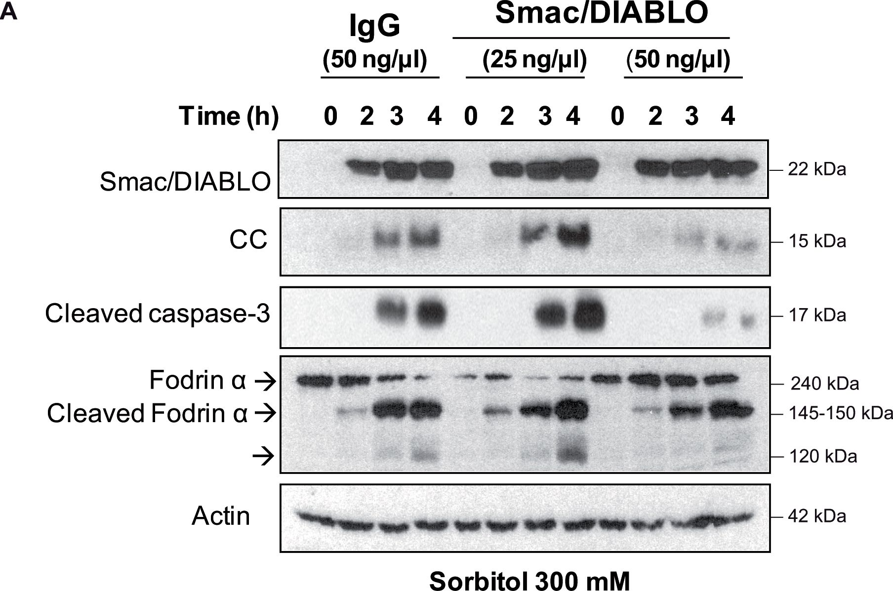
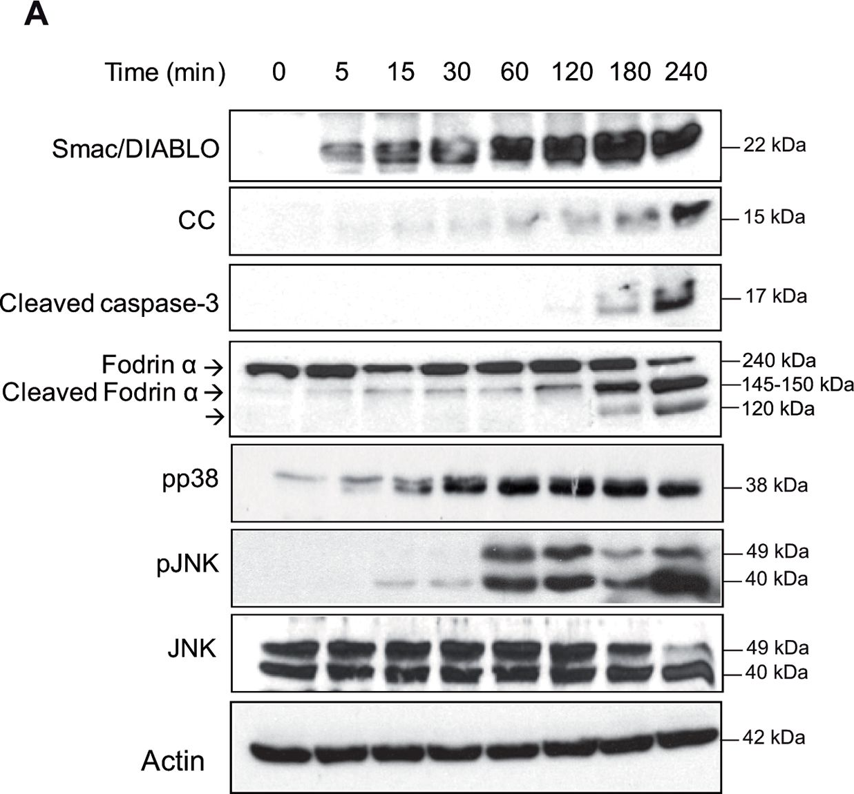
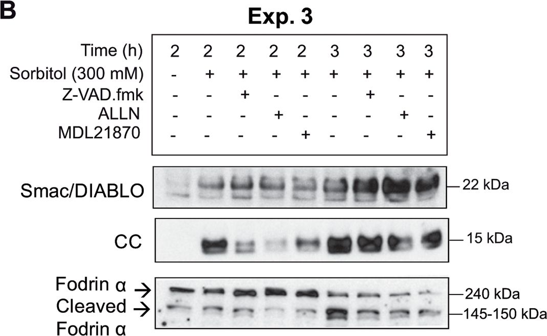
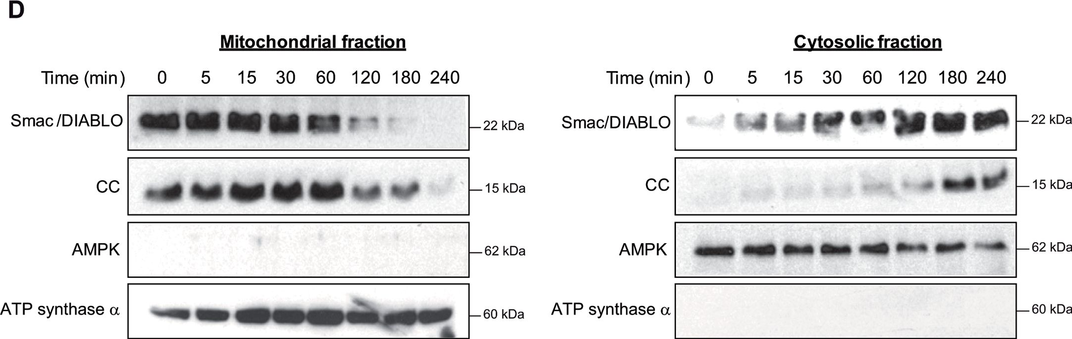
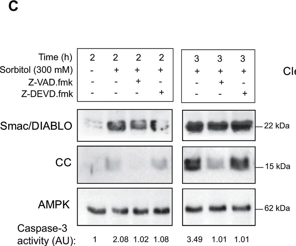
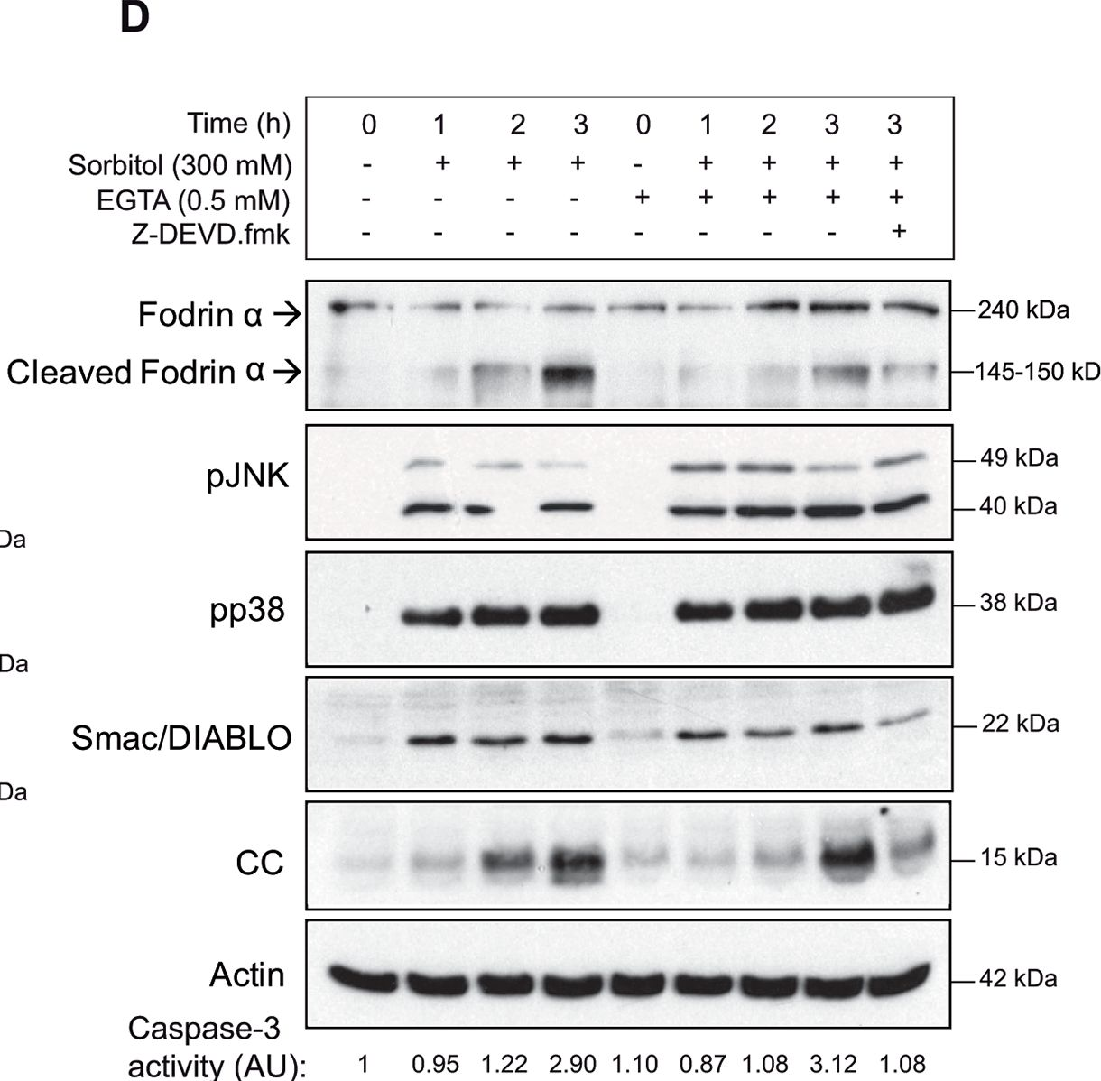
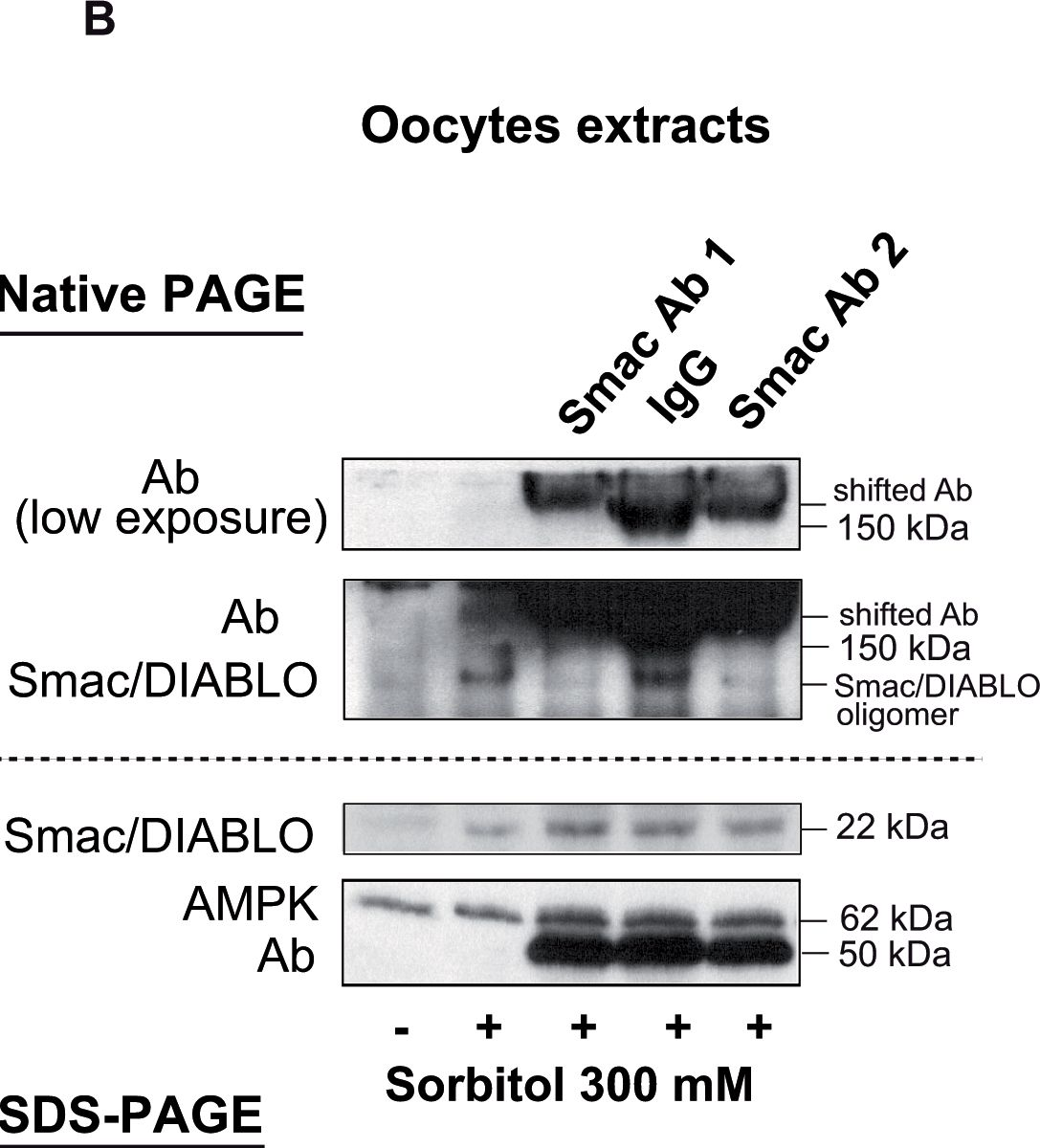
Hyperosmotic shock induces early calpain activation, Smac/DIABLO release from the mitochondria, and p38/JNK activation in Xenopus oocytes. These pathways regulate late cytochrome c release and caspase-3 activation. Here, we show that JNK1-1 and JNK1-2 are activated early by osmostress, and sustained activation of both isoforms accelerates the apoptotic program. When caspase-3 is activated, JNK1-2 is proteolyzed at Asp-385 increasing the release of cytochrome c and caspase-3 activity, thereby creating a positive feedback loop. Expression of Bcl-xL markedly reduces hyperosmotic shock-induced apoptosis. In contrast, expression of Bid induces rapid caspase-3 activation, even in the absence of osmostress, which is blocked by Bcl-xL co-expression. In these conditions a significant amount of Bid in the cytosol is mono- and bi-ubiquitinated. Caspase-3 activation by hyperosmotic shock induces proteolysis of Bid and mono-ubiquitinated Bid at Asp-52 increasing the release of cytochrome c and caspase-3 activation, and thus creating a second positive feedback loop. Revealing the JNK isoforms and the loops activated by osmostress could help to design better treatments for human diseases caused by perturbations in fluid osmolarity.© 2015 by The American Society for Biochemistry and Molecular Biology, Inc.
-
Biochemistry and Molecular biology
In Molecular Biology of the Cell on 15 December 2015 by Yamamori, T., Ike, S., et al.
Accumulating evidence suggests that mitochondrial dynamics is crucial for the maintenance of cellular quality control and function in response to various stresses. However, the role of mitochondrial dynamics in cellular responses to ionizing radiation (IR) is still largely unknown. In this study, we provide evidence that IR triggers mitochondrial fission mediated by the mitochondrial fission protein dynamin-related protein 1 (Drp1). We also show IR-induced mitotic catastrophe (MC), which is a type of cell death associated with defective mitosis, and aberrant centrosome amplification in mouse embryonic fibroblasts (MEFs). These are attenuated by genetic or pharmacological inhibition of Drp1. Whereas radiation-induced aberrant centrosome amplification and MC are suppressed by the inhibition of Plk1 and CDK2 in wild-type MEFs, the inhibition of these kinases is ineffective in Drp1-deficient MEFs. Furthermore, the cyclin B1 level after irradiation is significantly higher throughout the time course in Drp1-deficient MEFs than in wild-type MEFs, implying that Drp1 is involved in the regulation of cyclin B1 level. These findings strongly suggest that Drp1 plays an important role in determining the fate of cells after irradiation via the regulation of mitochondrial dynamics.
© 2015 Yamamori et al. This article is distributed by The American Society for Cell Biology under license from the author(s). Two months after publication it is available to the public under an Attribution–Noncommercial–Share Alike 3.0 Unported Creative Commons License (http://creativecommons.org/licenses/by-nc-sa/3.0).
-
Cell Biology
In PLoS One on 14 April 2015 by Ben Messaoud, N., Yue, J., et al.
Fig.3.A
-
WB
-
Xenopus laevis (African clawed frog)
Collected and cropped from PLoS ONE by CiteAb, provided under a CC-BY license
Image 1 of 8
In PLoS One on 14 April 2015 by Ben Messaoud, N., Yue, J., et al.
Fig.1.A
-
WB
-
Xenopus laevis (African clawed frog)
Collected and cropped from PLoS ONE by CiteAb, provided under a CC-BY license
Image 1 of 8
In PLoS One on 14 April 2015 by Ben Messaoud, N., Yue, J., et al.
Fig.2.B
-
WB
-
Xenopus laevis (African clawed frog)
Collected and cropped from PLoS ONE by CiteAb, provided under a CC-BY license
Image 1 of 8
In PLoS One on 14 April 2015 by Ben Messaoud, N., Yue, J., et al.
Fig.1.D
-
WB
-
Xenopus laevis (African clawed frog)
Collected and cropped from PLoS ONE by CiteAb, provided under a CC-BY license
Image 1 of 8
In PLoS One on 14 April 2015 by Ben Messaoud, N., Yue, J., et al.
Fig.2.C
-
WB
-
Xenopus laevis (African clawed frog)
Collected and cropped from PLoS ONE by CiteAb, provided under a CC-BY license
Image 1 of 8
In PLoS One on 14 April 2015 by Ben Messaoud, N., Yue, J., et al.
Fig.2.D
-
WB
-
Xenopus laevis (African clawed frog)
Collected and cropped from PLoS ONE by CiteAb, provided under a CC-BY license
Image 1 of 8
In PLoS One on 14 April 2015 by Ben Messaoud, N., Yue, J., et al.
Fig.3.B
-
WB
-
Xenopus laevis (African clawed frog)
Collected and cropped from PLoS ONE by CiteAb, provided under a CC-BY license
Image 1 of 8
In PLoS One on 14 April 2015 by Ben Messaoud, N., Yue, J., et al.
Fig.3.C

-
WB
-
Xenopus laevis (African clawed frog)
Collected and cropped from PLoS ONE by CiteAb, provided under a CC-BY license
Image 1 of 8