Tumor necrosis happens commonly in advanced solid tumors. We reported that necroptosis plays a major role in tumor necrosis. Although several key necroptosis regulators including receptor interacting protein kinase 1 (RIPK1) have been identified, the regulation of tumor necroptosis during tumor development remains elusive. Here, we report that Z-DNA-binding protein 1 (ZBP1), not RIPK1, mediates tumor necroptosis during tumor development in preclinical cancer models. We found that ZBP1 expression is dramatically elevated in necrotic tumors. Importantly, ZBP1, not RIPK1, deletion blocks tumor necroptosis during tumor development and inhibits metastasis. We showed that glucose deprivation triggers ZBP1-depedent necroptosis in tumor cells. Glucose deprivation causes mitochondrial DNA (mtDNA) release to the cytoplasm and the binding of mtDNA to ZBP1 to activate MLKL in a BCL-2 family protein, NOXA-dependent manner. Therefore, our study reveals ZBP1 as the key regulator of tumor necroptosis and provides a potential drug target for controlling tumor metastasis.
Product Citations: 19
Applications
Reactivity
Research Area
ZBP1 not RIPK1 mediates tumor necroptosis in breast cancer.
In Nature Communications on 11 May 2021 by Baik, J. Y., Liu, Z., et al.
-
Cancer Research
In Cell Death and Differentiation on 1 September 2019 by Tailler, M., Lindqvist, L. M., et al.
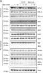
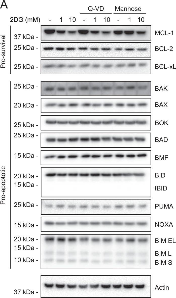
Drugs targeting various pro-survival BCL-2 family members (''BH3 mimetics'') have efficacy in hemopoietic malignancies, but the non-targeted pro-survival family members can promote resistance. Pertinently, the sensitivity of some tumor cell lines to BH3 mimetic ABT737, which targets BCL-2, BCL-XL, and BCL-W but not MCL-1, is enhanced by 2-deoxyglucose (2DG). We found that 2DG augmented apoptosis induced by ABT737 in 3 of 8 human hemopoietic tumor cell lines, most strongly in pre-B acute lymphocytic leukemia cell line NALM-6, the focus of our mechanistic studies. Although 2DG can lower MCL-1 translation, how it does so is incompletely understood, in part because 2DG inhibits both glycolysis and protein glycosylation in the endoplasmic reticulum (ER). Its glycolysis inhibition lowered ATP and, through the AMPK/mTORC1 pathway, markedly reduced global protein synthesis, as did an ER integrated stress response. A dual reporter assay revealed that 2DG impeded not only cap-dependent translation but also elongation or cap-independent translation. MCL-1 protein fell markedly, whereas 12 other BCL-2 family members were unaffected. We ascribe the MCL-1 drop to the global fall in translation, exacerbated for mRNAs with a structured 5' untranslated region (5'UTR) containing potential regulatory motifs like those in MCL-1 mRNA and the short half-life of MCL-1 protein. Pertinently, 2DG downregulated two other short-lived oncoproteins, MYC and MDM2. Thus, our results support MCL-1 as a critical 2DG target, but also reveal multiple effects on global translation that may well also affect its promotion of apoptosis.
-
WB
-
Homo sapiens (Human)
-
Biochemistry and Molecular biology
-
Cancer Research
-
Cell Biology
-
Genetics
In Cell Death and Differentiation on 1 July 2019 by Anstee, N. S., Bilardi, R. A., et al.
Many acute myeloid leukaemias (AMLs) express high levels of BCL-2 and MCL-1, especially after therapy. To test the impact of these anti-apoptotic proteins on AML development and treatment, we used haemopoietic reconstitution to generate MLL-AF9 AMLs expressing BCL-2 or Mcl-1 transgenes. AMLs with elevated BCL-2 or MCL-1 had a higher proportion of mature myeloid cells but, like conventional MLL-AF9 AMLs, were readily transplantable. Short-term cell lines established from multiple primary AMLs of each genotype were tested in vitro for susceptibility to chemotherapeutics currently used for treating AML (daunorubicin, etoposide, cytarabine); the proteasome inhibitor bortezomib; CDK7/9 inhibitors; and BH3 mimetics, which bind and inhibit pro-survival proteins. The BH3 mimetics tested, alone and in combination with the other drugs, were: ABT-737 which, like its clinical counterpart navitoclax, targets BCL-2, BCL-XL and BCL-W; BCL-2-specific ABT-199 (venetoclax); BCL-XL-specific A-1331852; and S63845, a new MCL-1-specific BH3 mimetic. As single agents, daunorubicin and bortezomib had the greatest efficacy. Elevated MCL-1 or BCL-2 reduced sensitivity to daunorubicin but, surprisingly, not to bortezomib. MCL-1 markedly enhanced resistance to ABT-737 and ABT-199 but not S63845, and BCL-2 increased resistance to S63845 but not to ABT-737 or ABT-199. Notable synergies were achieved by combining BH3 mimetics with daunorubicin: S63845 increased the sensitivity of both MCL-1 and BCL-2 overexpressing MLL-AF9 AMLs, and ABT-737 aided in killing those overexpressing BCL-2. Synergy between daunorubicin and ABT-199 was also apparent in vivo, although not curative. Impressive synergistic responses were achieved for human MLL-fusion AML cell lines treated with daunorubicin plus either ABT-737, ABT-199 or S63845, and with ABT-199 plus S63845, with or without daunorubicin. Our data suggest that AML patients may benefit from combining conventional cytotoxic drugs with BH3 mimetics targeting BCL-2 or MCL-1 or, if tolerated, both these agents.
-
Cell Biology
MicroRNA-711-Induced Downregulation of Angiopoietin-1 Mediates Neuronal Cell Death.
In Journal of Neurotrauma on 15 October 2018 by Sabirzhanov, B., Faden, A. I., et al.
Angiopoietin-1 (Ang-1) is a well-known endothelial growth factor, but its effects on neurons have yet to be elucidated. We show that Ang-1 is rapidly downregulated in the injured brain after controlled cortical impact (CCI), a mouse experimental traumatic brain injury (TBI) model and in etoposide-induced neuronal apoptosis in vitro. Ang-1 treatment inhibits etoposide-induced upregulation of proapoptotic B-cell lymphoma 2 (Bcl-2) family members Noxa, p53 upregulated modulator of apoptosis (Puma), Bcl-2 interacting mediator of cell death (Bim), and Bcl-2-associated X protein (Bax); reduces markers of caspase-dependent (cytochrome c release/caspase activation) and caspase-independent (apoptosis-inducing factor release) pathways; and limits neuronal cell death. Ang-1 treatment phosphorylates receptors Tunica interna endothelial cell kinase 2 (Tie2), and β1-integrin and limits the etoposide-induced decrease in protein kinase B (Akt) activity. Blocking Tie2 and β1-integrin signaling reduces Ang-1 neuroprotective effects. After both TBI and etoposide treatment microRNA (miR)-711 are upregulated, consistent with its putative role as a negative regulator of Ang-1. We show that miR-711 directly targets the Ang-1 messenger RNA (mRNA), decreasing Ang-1 expression. Increased levels of miR-711 and Ang-1 mRNA are found in the RNA-induced silencing complex complex site of miR-mediated degradation of target mRNAs after etoposide treatment and the miR-711mimic downregulates Ang-1. Administration of miR-711 inhibitor elevates Ang-1 after TBI whereas Ang-1 administration increases Akt activation; reduces Puma, Noxa, Bim, and Bax levels; and attenuates caspase-dependent and -independent neuronal apoptosis 24 h after TBI. Ang-1 also attenuates neuronal degeneration, increases gene expression of molecules that maintain blood-brain barrier integrity, and reduces post-traumatic lesion volume/edema 24 h after TBI. Although we only observed short-term neuroprotective effects after Ang-1 administration, miR-711-dependent downregulation of Ang-1, followed by Akt pathway inhibition, may play a role in neuronal cell death after neuronal injury in vitro and after experimental TBI.
In Frontiers in Cellular Neuroscience on 15 February 2018 by Duval, N., Sumner, W. A., et al.
The molecular mechanisms leading to motor neuron death in amyotrophic lateral sclerosis (ALS) are unknown; however, several studies have provided evidence of a central role for intrinsic apoptosis. Bcl-2 homology-3 domain (BH3)-only proteins are pro-apoptotic members of the Bcl-2 family whose enhanced expression acts as a trigger for the intrinsic apoptotic cascade. Here, we compared the relative expression of BH3-only proteins in the spinal cord of end-stage G93A mutant SOD1 mice to age-matched wild-type (WT) mice. Large alpha motor neurons in lumbar spinal cord sections of both WT and end-stage mutant SOD1 mice stained positively for a number of BH3-only proteins; however, no discernible differences were observed in either the relative intensity of staining or number of BH3-immunoreactive motor neurons between WT and mutant SOD1 mice. On the other hand, we observed significantly enhanced staining for Bid, DP5/Hrk, and BNip3L in GFAP-positive astrocytes only in end-stage G93A mutant SOD1 spinal cord. Staining of additional end-stage G93A mutant SOD1 tissues showed specific upregulation of DP5/Hrk in lumbar spinal cord sections, but not in cerebellum or cortex. Finally, examination of protein expression using western blotting also revealed marked increases in DP5/Hrk and BNip3L in G93A mutant SOD1 lumbar spinal cord lysates compared to WT controls. The upregulation of a specific subset of BH3-only proteins, including Bid, DP5/Hrk, and BNip3L, in reactive astrocytes suggests that these proteins may execute a novel non-apoptotic function within astrocytes to promote ALS disease progression, thus providing a new potential target for therapeutic intervention.
-
IHC
-
Mus musculus (House mouse)
-
Neuroscience
In Cell Death Differ on 1 September 2019 by Tailler, M., Lindqvist, L. M., et al.
Fig.3.A
-
WB
-
Homo sapiens (Human)
Collected and cropped from Cell Death and Differentiation by CiteAb, provided under a CC-BY license
Image 1 of 1