Transforming growth factor beta (TGFβ) and activin A suppress natural killer (NK) cell function and proliferation, limiting the efficacy of adoptive NK cell therapies. Inspired by the partial resistance to TGFβ of NK cells with SMAD4 haploinsufficiency, we used CRISPR-Cas9 for knockout of SMAD4 in human NK cells. Here we show that SMAD4KO NK cells were resistant to TGFβ and activin A inhibition, retaining their cytotoxicity, cytokine secretion and interleukin-2/interleukin-15-driven proliferation. They showed enhanced tumor penetration and tumor growth control, both as monotherapy and in combination with tumor-targeted therapeutic antibodies. Notably, SMAD4KO NK cells outperformed control NK cells treated with a TGFβ inhibitor, underscoring the benefit of maintaining SMAD4-independent TGFβ signaling. SMAD4KO conferred TGFβ resistance across diverse NK cell platforms, including CD19-CAR NK cells, stem cell-derived NK cells and ADAPT-NK cells. These findings position SMAD4 knockout as a versatile and compelling strategy to enhance NK cell antitumor activity, providing a new avenue for improving NK cell-based cancer immunotherapies.
© 2025. The Author(s).
Product Citations: 145
In Nature Immunology on 1 April 2025 by Rea, A., Santana-Hernández, S., et al.
-
Immunology and Microbiology
In Frontiers in Immunology on 27 September 2024 by Selim, M. A., Suef, R. A., et al.
The relationship between chronic hepatitis B (CHB) infection and natural killer (NK) cell dysfunction is well-established, but the specific role of HBV viral antigens in driving NK cell impairment in patients with CHB remains unclear. This study investigates the modulatory effects of hepatitis B virus subviral particles (HBVsvp, a representative model for HBsAg) on the phenotypic regulation (activating and inhibitory receptors), cytokine production and cytotoxic potential of peripheral blood mononuclear cell-derived natural killer cells (PBMCs-derived NK cell), which contributes to NK cell dysfunction in CHB infection, potentially serving as an effective HBV immune evasion strategy by the virus.
NK cells were isolated from peripheral blood of patients with CHB (n=5) and healthy individuals (n=5), stimulated with HBVsvp. Subsequent flow cytometric characterization involved assessing changes in activating (NKp46 and NKG2D) and inhibitory (CD94) receptors expression, quantifying TNF-α and IFN- γ cytokine secretion, and evaluating the cytotoxic response against HepG2.2.15 cells with subsequent HBVsvp quantification.
In CHB patients, in vitro exposure of PBMCs-derived NK cell with HBVsvp (represent HBsAg model) significantly reduced NK cell-activating receptors expression (P = 0.022), increased expression of CD94 + NK cells (p = 0.029), accompanied with a reduced TNF-α - IFN-γ cytokine levels, and impaired cytotoxic capacity (evidenced by increased cell proliferation and elevated HBVsvp levels in co-cultures with HepG2.2.15 cells in a time-dependent), relative to healthy donors.
These findings suggest that HBVsvp may induce dysfunctional NK cell responses characterized by phenotypic imbalance with subsequent reduction in cytokine and cytotoxic levels, indicating HBVsvp immunosuppressive effect that compromises antiviral defense in CHB patients. These data enhance our understanding of NK cell interactions with HBsAg and highlight the potential for targeting CD94 inhibitory receptors to restore NK cell function as an immunotherapeutic approach. Further clinical research is needed to validate these observations and establish their utility as reliable biomarkers.
Copyright © 2024 Selim, Suef, Saied, Abdel-Maksoud, Almutairi, Aufy, Mousa, Mansour and Farag.
-
Homo sapiens (Human)
-
Immunology and Microbiology
In Frontiers in Immunology on 11 July 2024 by Jorisch-Mühlebach, O., Pitts, D., et al.
Hemophagocytic lymphohistiocytosis (HLH) is a life-threatening immune disorder characterized by uncontrolled lymphocyte and macrophage activation and a subsequent cytokine storm. The timely initiation of immunosuppressive treatment is crucial for survival.
Here, we harnessed Vγ9Vδ2 T cell degranulation to develop a novel functional assay for the diagnosis of HLH. We compared the novel assay with the conventional natural killer (NK) cell stimulation method in terms of efficiency, specificity, and reliability. Our analysis involved 213 samples from 182 individuals, including 23 samples from 12 patients with degranulation deficiency (10 individuals with UNC13D deficiency, 1 with STXBP2 deficiency, and 1 with RAB27A deficiency).
While both tests exhibited 100% sensitivity, the Vγ9Vδ2 T cell degranulation assay showed a superior specificity of 86.2% (n=70) compared to the NK cell degranulation assay, which achieved 78.9% specificity (n=213). The Vγ9Vδ2 T cell degranulation assay offered simpler technical requirements and reduced labor intensity, leading to decreased susceptibility to errors with faster processing times.
This efficiency stemmed from the sole requirement of dissolving (E)-4-hydroxy-3-methyl-but-2-enyl pyrophosphate (HMBPP) powder, contrasting with the intricate maintenance of K562 cells necessary for the NK cell degranulation assay. With its diminished susceptibility to errors, we anticipate that the assay will require fewer repetitions of analysis, rendering it particularly well-suited for testing infants.
The Vγ9Vδ2 T cell degranulation assay is a user-friendly, efficient diagnostic tool for HLH. It offers greater specificity, reliability, and practicality than established methods. We believe that our present findings will facilitate the prompt, accurate diagnosis of HLH and thus enable rapid treatment and better patient outcomes.
Copyright © 2024 Jorisch-Mühlebach, Pitts, Tinner, Teh, Roelli, Prader, Vavassori and Pachlopnik Schmid.
-
Homo sapiens (Human)
-
Immunology and Microbiology
Preprint on MedRxiv : the Preprint Server for Health Sciences on 14 May 2024 by Natalini Martínez, S., Ramos, R., et al.
In this phase III, open label, single arm, multicenter clinical study, we report safety, tolerability and immunogenicity of PHH-1V as a booster dose in subjects primary vaccinated against COVID-19 with the BNT162b2, mRNA-1273, ChAdOx1-S, or Ad26.COV2.S vaccines, with or without previous COVID-19 infection. A total of 2661 subjects were included in this study and vaccinated with the PHH-1V vaccine. Most treatment-emergent adverse events (TEAE) were solicited local and systemic reactions with grade 1 (58.70%) or grade 2 (27.58%) intensity, being the most frequently reported injection site pain (82.83%), fatigue (31.72%) and headache (31.23%). Additionally, immunogenicity was assessed at Baseline and Days 14, 91, 182 and 365 in a subset of 235 subjects primary vaccinated. On Day 14, geometric mean triter (GMT) in neutralizing antibody against SARS-CoV-2 Wuhan and Beta, Delta and Omicron BA.1 variants increased in all primary vaccination with a geometric mean fold raise (GMFR) of 6.90 (95% CI 4.96-9.58), 12.27 (95% CI 8.52-17.67), 7.24 (95% CI 5.06-10.37) and 17.51 (95% CI 12.28-24.97), respectively. Despite GMT decay after day 14, it remains in all cases significatively higher from baseline up to 1 year after PHH-1V booster administration and GMFR against Beta and Omicron BA.1 variants over 3 at 1 year after booster compared to baseline. PHH-1V booster vaccination elicited also a significant RBD/Spike-specific IFN-γ + T-cell responses on Day 14. Overall, PHH-1V vaccine was immunogenic and well-tolerated regardless of the previous primary vaccination scheme received with no reported cases of severe COVID-19 infection throughout the entire study.
-
COVID-19
-
Immunology and Microbiology
In Bio-protocol on 5 May 2024 by Lim, H. J. & McWilliam, H. E. G.
Plasma membrane proteins mediate important aspects of physiology, including nutrient acquisition, cell-cell interactions, and monitoring homeostasis. The trafficking of these proteins, involving internalisation from and/or recycling back to the cell surface, is often critical to their functions. These processes can vary among different proteins and cell types and states and are still being elucidated. Current strategies to measure surface protein internalisation and recycling are typically microscopy or biochemical assays; these are accurate but generally limited to analysing a homogenous cell population and are often low throughput. Here, we present flow cytometry-based methods involving probe-conjugated antibodies that enable quantification of internalisation or recycling rates at the single-cell level in complex samples. To measure internalisation, we detail an assay where the protein of interest is labelled with a specific antibody conjugated to a fluorescent oligonucleotide-labelled probe. To measure recycling, a specific antibody conjugated to a cleavable biotin group is employed. These probes permit the differentiation of molecules that have been internalised or recycled from those that have not. When combined with cell-specific marker panels, these methods allow the quantitative study of plasma membrane protein trafficking dynamics in a heterogenous cell mixture at the single-cell level. Key features • These assays allow sensitive quantification of internalised or recycled surface molecules using oligonucleotide or cleavable biotin-conjugated probes, respectively, and detected by flow cytometry. • They can be adapted to any membrane protein that transits via the cell surface and for which a specific purified antibody is available. • The dynamics of a cell surface protein can be measured in heterogenous cell populations simultaneously, including various cellular activation states. • The internalisation assay builds upon the method developed by Liu et al. [1,2] and extends its application to heterogenous human peripheral blood mononuclear cells. • These assays have been extensively used on suspension cells but have not been tested on adherent cells.
©Copyright : © 2024 The Authors; This is an open access article under the CC BY-NC license.
-
Homo sapiens (Human)
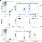
In Front Immunol on 20 August 2022 by Heropolitanska-Pliszka, E., Piątosa, B., et al.
Fig.2.B

-
FC/FACS
-
Collected and cropped from Front Immunol by CiteAb, provided under a CC-BY license
Image 1 of 1