Induction of durable protective immune responses is the main goal of prophylactic vaccines, and adjuvants play a role as drivers of such responses. Despite advances in vaccine strategies, development of a safe and effective HIV vaccine remains a significant challenge. Use of an appropriate adjuvant is crucial to the success of HIV vaccines. Here we assessed the saponin/MPLA nanoparticle (SMNP) adjuvant with an HIV envelope (Env) trimer, evaluating the safety and effect of multiple variables - including adjuvant dose (16-fold dose range), immunization route, and adjuvant composition - on the establishment of Env-specific memory T and B cell (TMem and BMem) responses and long-lived plasma cells in nonhuman primates (NHPs). Robust BMem were detected in all groups, but a 6-fold increase was observed in the highest- versus the lowest-SMNP-dose group. Similarly, stronger vaccine responses were induced by the highest SMNP dose in CD40L+OX40+ CD4+ TMem (11-fold), IFN-γ+ CD4+ TMem (15-fold), IL21+ CD4+ TMem (9-fold), circulating T follicular helper cells (TFH; 3.6-fold), BM plasma cells (7-fold), and binding IgG (1.3-fold). Substantial tier 2 neutralizing antibodies were only observed in the higher-SMNP-dose groups. These investigations highlight the dose-dependent potency of SMNP and its relevance for human use and next-generation vaccines.
Product Citations: 7
In The Journal of Clinical Investigation on 15 April 2025 by Ramezani-Rad, P., Marina-Zárate, E., et al.
-
FC/FACS
-
Immunology and Microbiology
In Journal of Nanobiotechnology on 29 January 2025 by Zhao, S., Kong, H., et al.
Hypertrophic scar (HS) is a common fibroproliferative disorders with no fully effective treatments. The conversion of fibroblasts to myofibroblasts is known to play a critical role in HS formation, making it essential to identify molecules that promote myofibroblast dedifferentiation and to elucidate their underlying mechanisms. In this study, we used comparative transcriptomics and single-cell sequencing to identify key molecules and pathways that mediate fibrosis and myofibroblast transdifferentiation. Epidermal stem cell-derived extracellular vesicles (EpiSC-EVs) were isolated via ultracentrifugation and filtration, followed by miRNA sequencing to identify miRNAs targeting key molecules. After in vitro and in vivo treatment with EpiSC-EVs, we assessed antifibrotic effects through scratch assays, collagen contraction assays, Western blotting, and immunofluorescence. Transcriptomic sequencing and rescue experiments were used to investigate the molecular mechanism by which miR-203a-3p in EpiSC-EVs induces myofibroblast dedifferentiation. Our results indicate that PIK3CA is overexpressed in HS tissues and positively correlates with fibrosis. EpiSC-EVs were absorbed by scar-derived fibroblasts, promoting dedifferentiation from myofibroblasts to quiescent fibroblasts. Mechanistically, miR-203a-3p in EpiSC-EVs plays an essential role in inhibiting PIK3CA expression and PI3K/AKT/mTOR pathway hyperactivation, thereby reducing scar formation. In vivo studies confirmed that EpiSC-EVs attenuate excessive scarring through the miR-203a-3p/PIK3CA axis, suggesting EpiSC-EVs as a promising therapeutic approach for HS.
© 2025. The Author(s).
-
FC/FACS
-
Stem Cells and Developmental Biology
Application of In vitro transcytosis models to brain targeted biologics.
In PLoS ONE on 23 August 2023 by Deng, K., Lu, Y., et al.
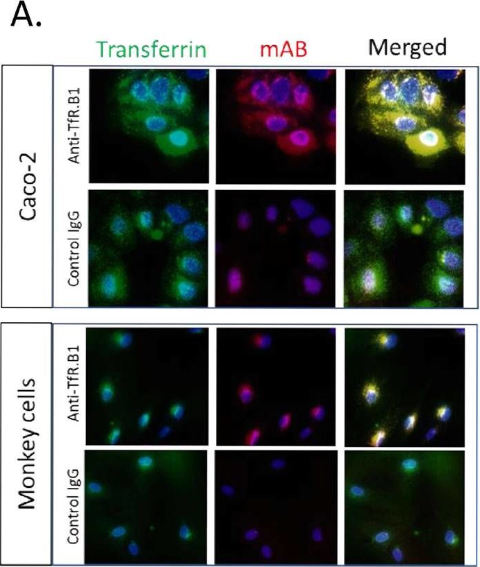
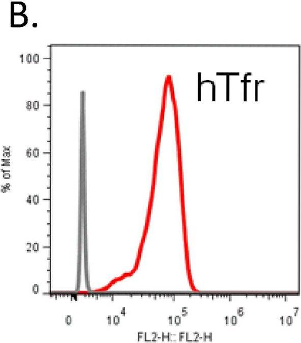
The blood brain barrier (BBB) efficiently limits the penetration of biologics drugs from blood to brain. Establishment of an in vitro BBB model can facilitate screening of central nervous system (CNS) drug candidates and accelerate CNS drug development. Despite many established in vitro models, their application to biologics drug selection has been limited. Here, we report the evaluation of in vitro transcytosis of anti-human transferrin receptor (TfR) antibodies across human, cynomolgus and mouse species. We first evaluated human models including human cerebral microvascular endothelial cell line hCMEC/D3 and human colon epithelial cell line Caco-2 models. hCMEC/D3 model displayed low trans-epithelial electrical resistance (TEER), strong paracellular transport, and similar transcytosis of anti-TfR and control antibodies. In contrast, the Caco-2 model displayed high TEER value and low paracellular transport. Anti-hTfR antibodies demonstrated up to 70-fold better transcytosis compared to control IgG. Transcytosis of anti-hTfR.B1 antibody in Caco-2 model was dose-dependent and saturated at 3 μg/mL. Enhanced transcytosis of anti-hTfR.B1 was also observed in a monkey brain endothelial cell based (MBT) model. Importantly, anti-hTfR.B1 showed relatively high brain radioactivity concentration in a non-human primate positron emission tomography study indicating that the in vitro transcytosis from both Caco-2 and MBT models aligns with in vivo brain exposure. Typically, brain exposure of CNS targeted biologics is evaluated in mice. However, antibodies, such as the anti-human TfR antibodies, do not cross-react with the mouse target. Therefore, validation of a mouse in vitro transcytosis model is needed to better understand the in vitro in vivo correlation. Here, we performed transcytosis of anti-mouse TfR antibodies in mouse brain endothelial cell-based models, bEnd3 and the murine intestinal epithelial cell line mIEC. There is a good correlation between in vitro transcytosis of anti-mTfR antibodies and bispecifics in mIEC model and their mouse brain uptake. These data strengthen our confidence in the predictive power of the in vitro transcytosis models. Both mouse and human in vitro models will serve as important screening assays for brain targeted biologics selection in CNS drug development.
Copyright: © 2023 Deng et al. This is an open access article distributed under the terms of the Creative Commons Attribution License, which permits unrestricted use, distribution, and reproduction in any medium, provided the original author and source are credited.
-
IA
-
Homo sapiens (Human)
TAF10 Interacts with the GATA1 Transcription Factor and Controls Mouse Erythropoiesis.
In Molecular and Cellular Biology on 1 June 2015 by Papadopoulos, P., Gutiérrez, L., et al.
The ordered assembly of a functional preinitiation complex (PIC), composed of general transcription factors (GTFs), is a prerequisite for the transcription of protein-coding genes by RNA polymerase II. TFIID, comprised of the TATA binding protein (TBP) and 13 TBP-associated factors (TAFs), is the GTF that is thought to recognize the promoter sequences allowing site-specific PIC assembly. Transcriptional cofactors, such as SAGA, are also necessary for tightly regulated transcription initiation. The contribution of the two TAF10-containing complexes (TFIID, SAGA) to erythropoiesis remains elusive. By ablating TAF10 specifically in erythroid cells in vivo, we observed a differentiation block accompanied by deregulated GATA1 target genes, including Gata1 itself, suggesting functional cross talk between GATA1 and TAF10. Additionally, we analyzed by mass spectrometry the composition of TFIID and SAGA complexes in mouse and human cells and found that their global integrity is maintained, with minor changes, during erythroid cell differentiation and development. In agreement with our functional data, we show that TAF10 interacts directly with GATA1 and that TAF10 is enriched on the GATA1 locus in human fetal erythroid cells. Thus, our findings demonstrate a cross talk between canonical TFIID and SAGA complexes and cell-specific transcription activators during development and differentiation.Copyright © 2015, American Society for Microbiology. All Rights Reserved.
-
ChIP
-
Biochemistry and Molecular biology
-
Cell Biology
In Blood on 1 September 2011 by Libregts, S. F., Gutiérrez, L., et al.
Anemia of chronic disease is a complication accompanying many inflammatory diseases. The proinflammatory cytokine IFN-γ has been implicated in this form of anemia, but the underlying mechanism remains unclear. Here we describe a novel mouse model for anemia of chronic disease, in which enhanced CD27-mediated costimulation strongly increases the formation of IFN-γ-producing effector T cells, leading to a progressive anemia. We demonstrate that the anemia in these mice is fully dependent on IFN-γ and that this cytokine reduces both the life span and the formation of red blood cells. Molecular analysis revealed that IFN-γ induces expression of the transcription factors of interferon regulatory factor-1 (IRF-1) and PU.1 in both murine and human erythroid precursors. We found that, on IFN-γ stimulation, IRF-1 binds to the promoter of SPI.1 (PU.1) and induces PU.1 expression, leading to inhibition of erythropoiesis. Notably, down-regulation of either IRF-1 or PU.1 expression is sufficient to overcome IFN-γ-induced inhibition of erythropoiesis. These findings reveal a molecular mechanism by which chronic exposure to IFN-γ induces anemia.
-
ChIP
-
Mus musculus (House mouse)
-
Cardiovascular biology
In PLoS One on 23 August 2023 by Deng, K., Lu, Y., et al.
Fig.3.A
-
IA
-
Homo sapiens (Human)
Collected and cropped from PLoS ONE by CiteAb, provided under a CC-BY license
Image 1 of 2
In PLoS One on 23 August 2023 by Deng, K., Lu, Y., et al.
Fig.2.B
-
IA
-
Homo sapiens (Human)
Collected and cropped from PLoS ONE by CiteAb, provided under a CC-BY license
Image 1 of 2