
Homologous recombination (HR) is crucial for the high-fidelity repair of DNA double-strand breaks (DSBs), ensuring the maintenance of genome stability. In this study, we show that FOXD3 interacts with poly (ADP-ribose) polymerase 1 (PARP1) and is recruited to DSBs in a PARP1-dependent manner. FOXD3 directly binds to the DSB repair protein MRE11 and promotes its recruitment to DSB sites, ensuring proper end resection. Inhibition of FOXD3 expression compromises HR-mediated DSB repair and chromosome stability and sensitizes cancer cells to ionizing radiation. Collectively, our findings demonstrate that FOXD3 promotes HR-mediated DSB repair and genome stability.
Product Citations: 1,423
In Acta Biochimica et Biophysica Sinica on 13 June 2025 by Xu, S., Zhang, J., et al.
-
Genetics
Show More
Show Less
The class II myosin MYH4 safeguards genome integrity and suppresses tumor progression.
In The Journal of Clinical Investigation on 2 June 2025 by Thatte, J., Moisés da Silva, A., et al.
Loss-of-function mutations in genome maintenance genes fuel tumorigenesis through increased genomic instability. A subset of these tumor suppressors are challenging to identify due to context dependency, including functional interactions with other genes and pathways. Here, we searched for potential causal genes that impact tumor development and/or progression in breast cancer through functional-genetic screening of candidate genes. MYH4, encoding a class II myosin, emerged as a top hit impacting genomic stability. We show that MYH4 suppresses DNA replication stress by promoting replication licensing and replication fork progression. Moreover, we observed a strong synergistic relationship among class II myosins in suppressing replication-associated DNA damage. Genomic analysis of Pan-Cancer Analysis of Whole Genomes project breast cancer samples revealed frequent concomitant loss of TP53 with MYH4 and class II myosins on chromosome 17p. Notably, Myh4 disruption accelerated mouse mammary tumorigenesis in a Trp53-deficient background. In conclusion, our results suggest an unanticipated function of MYH4 in p53-mediated tumor suppression that can explain their combined loss in breast cancer.
-
Cancer Research
Show More
Show Less
Multigenerational cell tracking of DNA replication and heritable DNA damage.
In Nature on 1 June 2025 by Panagopoulos, A., Stout, M., et al.
Cell heterogeneity is a universal feature of life. Although biological processes affected by cell-to-cell variation are manifold, from developmental plasticity to tumour heterogeneity and differential drug responses, the sources of cell heterogeneity remain largely unclear1,2. Mutational and epigenetic signatures from cancer (epi)genomics are powerful for deducing processes that shaped cancer genome evolution3-5. However, retrospective analyses face difficulties in resolving how cellular heterogeneity emerges and is propagated to subsequent cell generations. Here, we used multigenerational single-cell tracking based on endogenously labelled proteins and custom-designed computational tools to elucidate how oncogenic perturbations induce sister cell asymmetry and phenotypic heterogeneity. Dual CRISPR-based genome editing enabled simultaneous tracking of DNA replication patterns and heritable endogenous DNA lesions. Cell lineage trees of up to four generations were tracked in asynchronously growing cells, and time-resolved lineage analyses were combined with end-point measurements of cell cycle and DNA damage markers through iterative staining. Besides revealing replication and repair dynamics, damage inheritance and emergence of sister cell heterogeneity across multiple cell generations, through combination with single-cell transcriptomics, we delineate how common oncogenic events trigger multiple routes towards polyploidization with distinct outcomes for genome integrity. Our study provides a framework to dissect phenotypic plasticity at the single-cell level and sheds light onto cellular processes that may resemble early events during cancer development.
© 2025. The Author(s).
-
Genetics
Show More
Show Less
In Cell Death and Differentiation on 29 May 2025 by Modafferi, S., Farina, S., et al.
Formation of cytoplasmic inclusions (CIs) of TDP-43 and FUS, along with DNA damage accumulation, is a hallmark of affected motor neurons in Amyotrophic Lateral Sclerosis (ALS). However, the impact of CIs on DNA damage response (DDR) and repair in this pathology remains unprobed. Here, we show that CIs of TDP-43 and FUSP525L, co-localizing with stress granules, lead to a dysfunctional DDR activation associated with physical DNA breakage. Inhibition of the activity of the DDR kinase ATM, but not of ATR, abolishes DDR signaling, indicating that DNA double-strand breaks (DSBs) are the primary source of DDR activation. In addition, cells with TDP-43 and FUSP525L CIs exhibit reduced DNA damage-induced RNA synthesis at DSBs. We previously showed that the two endoribonucleases DROSHA and DICER, also known to interact with TDP-43 and FUS during small RNA processing, contribute to DDR signaling at DSBs. Treatment with enoxacin, which stimulates DDR and repair by boosting the enzymatic activity of DICER, restores a proficient DDR and reduces DNA damage accumulation in cultured cells with CIs and in vivo in a murine model of ALS. In Drosophila melanogaster, Dicer-2 overexpression rescues TDP-43-mediated retinal degeneration. In summary, our results indicate that the harmful effects caused by TDP-43 and FUS CIs include genotoxic stress and that the pharmacological stimulation of the DNA damage signaling and repair counteracts it.
© 2025. The Author(s).
-
Cell Biology
-
Genetics
Show More
Show Less
In Zoological Research on 18 May 2025 by Huang, X. Y., Liu, X. Y., et al.
The DNA replication stress (RS) response is crucial for maintaining cellular homeostasis and promoting physiological longevity. However, the mechanisms by which long-lived species, such as bats, regulate RS to maintain genomic stability remain unclear. Also, recent studies have uncovered noncanonical roles of ribosome-associated factors in maintaining genomic stability. In this study, somatic skin fibroblasts from the long-lived big-footed bat ( Myotis pilosus) were examined, with results showing that bat cells exhibited enhanced RS tolerance compared to mouse cells. Comparative transcriptome analysis under RS conditions revealed pronounced species-specific transcriptional differences, including robust up-regulation of ribosome biogenesis genes in bat cells and a markedly reduced activation of the P53 signaling pathway. These features emphasize a distinct homeostatic strategy in bat cells. Nuclear fragile X mental retardation-interacting protein 1 ( Nufip1), a ribosome-associated factor highly expressed in bat fibroblasts, was identified as a potential integrator of ribosomal and P53 signaling via its association with ribosomal protein S27-like (Rps27l). These findings provide direct cellular and molecular evidence for a noncanonical RS response in bats, highlighting a deeper understanding of the biological characteristics and genomic maintenance mechanisms of long-lived species.
-
Genetics
Show More
Show Less
In Nat Commun on 30 November 2023 by Jaiswal, R. K., Lei, K. H., et al.
Fig.2.C

-
WB
-
S96 in STN1 IDR is essential for antagonizing MRE11-mediated degradation of nascent strand DNA and for protecting genome stability.A Sequence alignment showing that S96 is conserved in higher eukaryotes. S96 is marked in red. B SIRF detection of S96A, S96D, and WT-STN1 at normal and stalled forks...
more
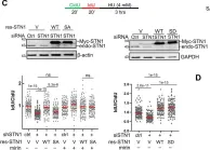
S96 in STN1 IDR is essential for antagonizing MRE11-mediated degradation of nascent strand DNA and for protecting genome stability.A Sequence alignment showing that S96 is conserved in higher eukaryotes. S96 is marked in red. B SIRF detection of S96A, S96D, and WT-STN1 at normal and stalled forks. Myc-tagged WT, S96A, S96D were stably expressed with retroviral transduction in U2OS cells treated with 4 mM HU for 3 h. Scale bars: 10 µm. Images with the red channel are provided in (Supplementary Fig. 9). Three independent experiments were performed and the result from one experiment is shown. P: One-way ANOVA. Red line: mean. n = 150 cells were analyzed per sample in each experiment. Western blot shows the expression of S96A, S96D, and WT-STN1. C DNA fiber analysis detecting NSD in the same U2OS cells described in (B). Three independent experiments were performed and the result from one experiment is shown. P: One-way ANOVA. Red line: mean. n = 100 fibers were measured per sample in each experiment. D DNA fiber analysis of NSD in BJ/hTERT cells co-expressing shSTN1 and RNAi-resistant S96A, S96D, and WT-STN1, which were expressed using retroviral transduction. Two independent experiments were performed and the result from one experiment is shown. P: One-way ANOVA. Red line: mean. n = 200 fibers were measured per sample in each experiment. E SIRF detection of MRE11 at normal and stalled replication forks in the same U2OS cells described in (B). Cells were pulse labeled with EdU for 8 min and treated with or without 4 mM HU for 3 h. Scale bars: 10 µm. Images with the red channel are provided in Supplementary Fig. 10. Three independent experiments were performed. P: One-way ANOVA. Red line: mean. Western blot showing expression of RNAi-resistant S96A, S96D, and WT-STN1. n = ~200 cells were analyzed per sample in each experiment. F Images showing chromosome aberrations in STN1 knockdown HeLa cells with ectopic expression of S96A, S96D, and WT-STN1. Cells were treated with HU (2 mM, 3 h). Aberrations are labeled with red stars. Examples of aberrant chromosomes are amplified and shown in inserts, with red arrows pointing to aberrations. Two independent experiments were performed, and the result from one experiment is shown. P: One-way ANOVA. Source data are provided in the Source Data file.
less
Collected and cropped from Nat Commun by CiteAb, provided under a CC-BY license
Image 1 of 29
In Nat Commun on 30 November 2023 by Jaiswal, R. K., Lei, K. H., et al.
Fig.1.B

-
WB
-
The STN1 IDR in the OB-fold domain protects against NSD under replication stress.A Top: Structure of the STN1-OB domain (PDB: 4JOI). PyMOL software shows that RPA32-OB/RPA14 and STN1-OB/TEN1 superimpose upon each other except the 26 aa (90–116) IDR present in STN1-OB. Bottom: NetSurfP-2.0 softwar...
more
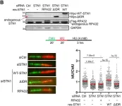
The STN1 IDR in the OB-fold domain protects against NSD under replication stress.A Top: Structure of the STN1-OB domain (PDB: 4JOI). PyMOL software shows that RPA32-OB/RPA14 and STN1-OB/TEN1 superimpose upon each other except the 26 aa (90–116) IDR present in STN1-OB. Bottom: NetSurfP-2.0 software predicts that STN1 90–116 is an IDR. The STN1-OB domain (aa 1–160) was used in prediction. B DNA fiber analysis of NSD in U2OS cells expressing RPA32, WT-STN1, or STN1-ΔIDR with concurrent knockdown of endogenous STN1. Flag-RPA32, Myc-WT-STN1, and Myc-STN1-ΔIDR were stably expressed by retroviral transduction. Three independent experiments were performed and the result from one experiment is shown. P: One-way ANOVA. The mean values are shown in red lines. n = 200 fibers were measured per sample in each experiment. Western blot shows STN1 knockdown and the expression of Flag-RPA32, Myc-WT-STN1, and Myc-STN1-ΔIDR. The expression level of endogenous STN1 is low (pointed by a red arrow). Note that Myc-STN1-ΔIDR migrates at almost the same position as the endogenous STN1. C Scheme of SIRF assay. Nascent strand DNA was pulse labeled with EdU to incorporate EdU at forks. Click chemistry was performed to covalently link biotin to EdU. Following incubation with primary antibodies (anti-biotin and anti-pS96) and secondary antibodies, PLA amplification was performed to visualize the proximity of phosphorylated STN1 to EdU-labeled forks. The scheme was created with BioRender.com. D SIRF detection of WT-STN1 and ΔIDR at normal and stalled forks. Myc-tagged WT-STN1 and ΔIDR were stably expressed in U2OS cells with retroviral transduction. Cells were pulse labeled with EdU for 8 min, then treated with or without HU (4 mM) for 3 h. Scale bars: 10 µm. Images with the red channel are provided in Supplementary Fig. 8. Two independent experiments were performed and the result from one experiment is shown. P: One-way ANOVA. Red line: mean. n = ~100 cells were measured per sample in each experiment. Source data are provided in the Source Data file.
less
Collected and cropped from Nat Commun by CiteAb, provided under a CC-BY license
Image 1 of 29
In Commun Biol on 2 March 2023 by Maresca, C., Dello Stritto, A., et al.
Fig.1.A

-
FC/FACS
-
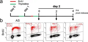
TRF1 and PARP1 interact during S-phase.HeLa cells were synchronized in the early S phase by double thymidine block. A BrdU pulse was administered 15 minutes before the second thymidine block and then the cells were released and collected at the indicated time points. a At each endpoint, the DNA w...
more
TRF1 and PARP1 interact during S-phase.HeLa cells were synchronized in the early S phase by double thymidine block. A BrdU pulse was administered 15 minutes before the second thymidine block and then the cells were released and collected at the indicated time points. a At each endpoint, the DNA was denatured, incubated with anti-BrdU antibody and PI, and processed for flow cytometry. Bivariate distributions (dot plot) of BrdU content versus DNA content (b) were analyzed, boundaries between positive and negative samples have been indicated as a black line (one representative of three independent experiments is shown). Histograms report the quantification of the percentage of cells in the different cell cycle phases in the whole cell population (c) or in BrdU pulsed population (d, the mean of three independent experiments is shown, bars are SD), 1C. e Samples synchronized as in a underwent immunoprecipitation with an anti-TRF1 specific antibody or rabbit IgG as negative control followed by incubation with the indicated antibodies (β-actin was used as loading control, one representative of three independent experiments is shown). Western blot signals were quantified by densitometry and reported in histograms after normalization on IP-ed TRF1, and on input PARP1 and TRF1, background in IgG IP-ed samples was subtracted (the mean of three independent experiments is reported, bars are SD). f HeLa cells synchronized as described were fixed in formaldehyde and processed for PLA with anti-TRF1 and PARP1 antibodies. Signals were acquired by Leica Deconvolution fluorescence microscope at 63× magnification (representative images are shown). The number of signals/nuclei was scored by Image J software and reported in graph (g). For each column Mean (red bars) and numerosity (N) are indicated, two pulled independent experiments were plotted, P value was determined by unpaired two tailed t-student test, ***P ≤ 0.001, ****P ≤ 0.0001.
less
Collected and cropped from Commun Biol by CiteAb, provided under a CC-BY license
Image 1 of 29
In Elife on 25 August 2020 by Hasenpusch-Theil, K., Laclef, C., et al.
Fig.3.E

-
IHC-IF
-
Mus musculus (House mouse)
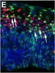
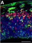
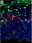
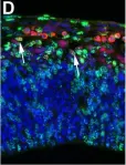
Increased neurogenesis at the expense of basal progenitor formation in the E12.5 Inpp5eΔ/Δ mutant lateral telencephalon.Immunohistochemistry on sections of E12.5 control (A, D) and Inpp5eΔ/Δ embryos (B, E) that were treated with BrdU 24 hr earlier. (A–C) Tbr2/BrdU double labeling showed that less...
more
Increased neurogenesis at the expense of basal progenitor formation in the E12.5 Inpp5eΔ/Δ mutant lateral telencephalon.Immunohistochemistry on sections of E12.5 control (A, D) and Inpp5eΔ/Δ embryos (B, E) that were treated with BrdU 24 hr earlier. (A–C) Tbr2/BrdU double labeling showed that less basal progenitors formed from the BrdU-labeled progenitor cohort in Inpp5eΔ/Δ embryos. (D–F) The proportion of newly formed Tbr1+ neurons was increased in Inpp5eΔ/Δ embryos. The arrows in D and E label Tbr1+BrdU+ cells. All statistical data are presented as means ± 95% confidence intervals (CI); unpaired t tests; n = 4; *p<0.05; **p<0.01. Scale bar: 50 μm.Apoptosis in the developing forebrain of Inpp5eΔ/Δ embryos.(A–F) Immunofluorescence staining for Cleaved Caspase three revealing apoptic cells. (A, B) Programmed cell death was detected in the midline roof plate (rp) but hardly in the developing neocortex of E12.5 control (A) and Inpp5eΔ/Δ embryos (B). (C–F) In E14.5 embryos, very few apoptotic cells were identified in the neocortex while apoptosis is widespread in the trigeminal ganglion (TG) (E, F). ctx: cortex. Scale bars: (A): 100 μm, (C): 25`0 μm, (D) 500 μm.Proportion of mitotic progenitors in Inpp5eΔ/Δ embryos.(A–H) Proportions of mitotic progenitors in E12.5 control (A, E) and Inpp5eΔ/Δ embryos (B, F) as revealed by pHH3 (mitotic cells) and PCNA (all progenitor cells) double immunofluorescence. Note the reduction in mitotic basal progenitors in the Inpp5eΔ/Δ medial neocortex (A, B, D). (I–P) The proportions of apical and basal progenitors is not significantly different in E14.5 control and Inpp5eΔ/Δ embryos. In all panels, radial glia cells divide at the ventricular surface, whereas mitotic basal progenitors locate in abventricular positions. All statistical data are presented as means ± 95% confidence intervals (CI); unpaired t-tests (C, G, K, L, P) and Mann Whitney tests (D, H, O); n = 4; *p<0.05. Scale bar: 50 μm.Cell cycle of cortical progenitors in E12.5 Inpp5eΔ/Δ embryos.(A) Schematic illustrating the BrdU/IdU double labeling strategy to measure S phase (TS) and total cell cycle length (TC). 90 min after an initial IdU administration, pregnant females received an intraperitoneal BrdU injection. Embryos are harvested 30 min later. (B, C) Double immunofluorescence to detect IdU+ and BrdU+ progenitors. (D) Quantification showing no significant change in TS and TC. Statistical data are presented as means ± 95% confidence intervals (CI); unpaired t-tests; n = 4; *p<0.05. Scale bar: 50 μm.
less
Collected and cropped from Elife by CiteAb, provided under a CC-BY license
Image 1 of 29
In Elife on 25 August 2020 by Hasenpusch-Theil, K., Laclef, C., et al.
Fig.3.A

-
IHC-IF
-
Mus musculus (House mouse)
Increased neurogenesis at the expense of basal progenitor formation in the E12.5 Inpp5eΔ/Δ mutant lateral telencephalon.Immunohistochemistry on sections of E12.5 control (A, D) and Inpp5eΔ/Δ embryos (B, E) that were treated with BrdU 24 hr earlier. (A–C) Tbr2/BrdU double labeling showed that less...
more
Increased neurogenesis at the expense of basal progenitor formation in the E12.5 Inpp5eΔ/Δ mutant lateral telencephalon.Immunohistochemistry on sections of E12.5 control (A, D) and Inpp5eΔ/Δ embryos (B, E) that were treated with BrdU 24 hr earlier. (A–C) Tbr2/BrdU double labeling showed that less basal progenitors formed from the BrdU-labeled progenitor cohort in Inpp5eΔ/Δ embryos. (D–F) The proportion of newly formed Tbr1+ neurons was increased in Inpp5eΔ/Δ embryos. The arrows in D and E label Tbr1+BrdU+ cells. All statistical data are presented as means ± 95% confidence intervals (CI); unpaired t tests; n = 4; *p<0.05; **p<0.01. Scale bar: 50 μm.Apoptosis in the developing forebrain of Inpp5eΔ/Δ embryos.(A–F) Immunofluorescence staining for Cleaved Caspase three revealing apoptic cells. (A, B) Programmed cell death was detected in the midline roof plate (rp) but hardly in the developing neocortex of E12.5 control (A) and Inpp5eΔ/Δ embryos (B). (C–F) In E14.5 embryos, very few apoptotic cells were identified in the neocortex while apoptosis is widespread in the trigeminal ganglion (TG) (E, F). ctx: cortex. Scale bars: (A): 100 μm, (C): 25`0 μm, (D) 500 μm.Proportion of mitotic progenitors in Inpp5eΔ/Δ embryos.(A–H) Proportions of mitotic progenitors in E12.5 control (A, E) and Inpp5eΔ/Δ embryos (B, F) as revealed by pHH3 (mitotic cells) and PCNA (all progenitor cells) double immunofluorescence. Note the reduction in mitotic basal progenitors in the Inpp5eΔ/Δ medial neocortex (A, B, D). (I–P) The proportions of apical and basal progenitors is not significantly different in E14.5 control and Inpp5eΔ/Δ embryos. In all panels, radial glia cells divide at the ventricular surface, whereas mitotic basal progenitors locate in abventricular positions. All statistical data are presented as means ± 95% confidence intervals (CI); unpaired t-tests (C, G, K, L, P) and Mann Whitney tests (D, H, O); n = 4; *p<0.05. Scale bar: 50 μm.Cell cycle of cortical progenitors in E12.5 Inpp5eΔ/Δ embryos.(A) Schematic illustrating the BrdU/IdU double labeling strategy to measure S phase (TS) and total cell cycle length (TC). 90 min after an initial IdU administration, pregnant females received an intraperitoneal BrdU injection. Embryos are harvested 30 min later. (B, C) Double immunofluorescence to detect IdU+ and BrdU+ progenitors. (D) Quantification showing no significant change in TS and TC. Statistical data are presented as means ± 95% confidence intervals (CI); unpaired t-tests; n = 4; *p<0.05. Scale bar: 50 μm.
less
Collected and cropped from Elife by CiteAb, provided under a CC-BY license
Image 1 of 29
In Elife on 25 August 2020 by Hasenpusch-Theil, K., Laclef, C., et al.
Fig.3.B

-
IHC-IF
-
Mus musculus (House mouse)
Increased neurogenesis at the expense of basal progenitor formation in the E12.5 Inpp5eΔ/Δ mutant lateral telencephalon.Immunohistochemistry on sections of E12.5 control (A, D) and Inpp5eΔ/Δ embryos (B, E) that were treated with BrdU 24 hr earlier. (A–C) Tbr2/BrdU double labeling showed that less...
more
Increased neurogenesis at the expense of basal progenitor formation in the E12.5 Inpp5eΔ/Δ mutant lateral telencephalon.Immunohistochemistry on sections of E12.5 control (A, D) and Inpp5eΔ/Δ embryos (B, E) that were treated with BrdU 24 hr earlier. (A–C) Tbr2/BrdU double labeling showed that less basal progenitors formed from the BrdU-labeled progenitor cohort in Inpp5eΔ/Δ embryos. (D–F) The proportion of newly formed Tbr1+ neurons was increased in Inpp5eΔ/Δ embryos. The arrows in D and E label Tbr1+BrdU+ cells. All statistical data are presented as means ± 95% confidence intervals (CI); unpaired t tests; n = 4; *p<0.05; **p<0.01. Scale bar: 50 μm.Apoptosis in the developing forebrain of Inpp5eΔ/Δ embryos.(A–F) Immunofluorescence staining for Cleaved Caspase three revealing apoptic cells. (A, B) Programmed cell death was detected in the midline roof plate (rp) but hardly in the developing neocortex of E12.5 control (A) and Inpp5eΔ/Δ embryos (B). (C–F) In E14.5 embryos, very few apoptotic cells were identified in the neocortex while apoptosis is widespread in the trigeminal ganglion (TG) (E, F). ctx: cortex. Scale bars: (A): 100 μm, (C): 25`0 μm, (D) 500 μm.Proportion of mitotic progenitors in Inpp5eΔ/Δ embryos.(A–H) Proportions of mitotic progenitors in E12.5 control (A, E) and Inpp5eΔ/Δ embryos (B, F) as revealed by pHH3 (mitotic cells) and PCNA (all progenitor cells) double immunofluorescence. Note the reduction in mitotic basal progenitors in the Inpp5eΔ/Δ medial neocortex (A, B, D). (I–P) The proportions of apical and basal progenitors is not significantly different in E14.5 control and Inpp5eΔ/Δ embryos. In all panels, radial glia cells divide at the ventricular surface, whereas mitotic basal progenitors locate in abventricular positions. All statistical data are presented as means ± 95% confidence intervals (CI); unpaired t-tests (C, G, K, L, P) and Mann Whitney tests (D, H, O); n = 4; *p<0.05. Scale bar: 50 μm.Cell cycle of cortical progenitors in E12.5 Inpp5eΔ/Δ embryos.(A) Schematic illustrating the BrdU/IdU double labeling strategy to measure S phase (TS) and total cell cycle length (TC). 90 min after an initial IdU administration, pregnant females received an intraperitoneal BrdU injection. Embryos are harvested 30 min later. (B, C) Double immunofluorescence to detect IdU+ and BrdU+ progenitors. (D) Quantification showing no significant change in TS and TC. Statistical data are presented as means ± 95% confidence intervals (CI); unpaired t-tests; n = 4; *p<0.05. Scale bar: 50 μm.
less
Collected and cropped from Elife by CiteAb, provided under a CC-BY license
Image 1 of 29
In Elife on 25 August 2020 by Hasenpusch-Theil, K., Laclef, C., et al.
Fig.3.D

-
IHC-IF
-
Mus musculus (House mouse)
Increased neurogenesis at the expense of basal progenitor formation in the E12.5 Inpp5eΔ/Δ mutant lateral telencephalon.Immunohistochemistry on sections of E12.5 control (A, D) and Inpp5eΔ/Δ embryos (B, E) that were treated with BrdU 24 hr earlier. (A–C) Tbr2/BrdU double labeling showed that less...
more
Increased neurogenesis at the expense of basal progenitor formation in the E12.5 Inpp5eΔ/Δ mutant lateral telencephalon.Immunohistochemistry on sections of E12.5 control (A, D) and Inpp5eΔ/Δ embryos (B, E) that were treated with BrdU 24 hr earlier. (A–C) Tbr2/BrdU double labeling showed that less basal progenitors formed from the BrdU-labeled progenitor cohort in Inpp5eΔ/Δ embryos. (D–F) The proportion of newly formed Tbr1+ neurons was increased in Inpp5eΔ/Δ embryos. The arrows in D and E label Tbr1+BrdU+ cells. All statistical data are presented as means ± 95% confidence intervals (CI); unpaired t tests; n = 4; *p<0.05; **p<0.01. Scale bar: 50 μm.Apoptosis in the developing forebrain of Inpp5eΔ/Δ embryos.(A–F) Immunofluorescence staining for Cleaved Caspase three revealing apoptic cells. (A, B) Programmed cell death was detected in the midline roof plate (rp) but hardly in the developing neocortex of E12.5 control (A) and Inpp5eΔ/Δ embryos (B). (C–F) In E14.5 embryos, very few apoptotic cells were identified in the neocortex while apoptosis is widespread in the trigeminal ganglion (TG) (E, F). ctx: cortex. Scale bars: (A): 100 μm, (C): 25`0 μm, (D) 500 μm.Proportion of mitotic progenitors in Inpp5eΔ/Δ embryos.(A–H) Proportions of mitotic progenitors in E12.5 control (A, E) and Inpp5eΔ/Δ embryos (B, F) as revealed by pHH3 (mitotic cells) and PCNA (all progenitor cells) double immunofluorescence. Note the reduction in mitotic basal progenitors in the Inpp5eΔ/Δ medial neocortex (A, B, D). (I–P) The proportions of apical and basal progenitors is not significantly different in E14.5 control and Inpp5eΔ/Δ embryos. In all panels, radial glia cells divide at the ventricular surface, whereas mitotic basal progenitors locate in abventricular positions. All statistical data are presented as means ± 95% confidence intervals (CI); unpaired t-tests (C, G, K, L, P) and Mann Whitney tests (D, H, O); n = 4; *p<0.05. Scale bar: 50 μm.Cell cycle of cortical progenitors in E12.5 Inpp5eΔ/Δ embryos.(A) Schematic illustrating the BrdU/IdU double labeling strategy to measure S phase (TS) and total cell cycle length (TC). 90 min after an initial IdU administration, pregnant females received an intraperitoneal BrdU injection. Embryos are harvested 30 min later. (B, C) Double immunofluorescence to detect IdU+ and BrdU+ progenitors. (D) Quantification showing no significant change in TS and TC. Statistical data are presented as means ± 95% confidence intervals (CI); unpaired t-tests; n = 4; *p<0.05. Scale bar: 50 μm.
less
Collected and cropped from Elife by CiteAb, provided under a CC-BY license
Image 1 of 29
In Nat Commun on 13 December 2019 by Brison, O., El-Hilali, S., et al.
Fig.4.C

-
ICC-IF
-
Homo sapiens (Human)
Short-term inhibition of transcription by triptolide does not impact CFS instability.a Determination of the minimal time permitting clearing of the FHIT gene from ongoing transcription. Upper panel: scheme of the experiments. Untreated cells (NT) and cells treated with 1 μM triptolide (Tpl) for t...
more
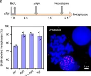
Short-term inhibition of transcription by triptolide does not impact CFS instability.a Determination of the minimal time permitting clearing of the FHIT gene from ongoing transcription. Upper panel: scheme of the experiments. Untreated cells (NT) and cells treated with 1 μM triptolide (Tpl) for the indicated periods of time were pulse labelled with EU for 30 min before recovery. Nascent RNAs were prepared by the Click-It method and quantified by RT-qPCR. Middle panel: map of the human FHIT gene with the position of exons (e1–e10), T-SDW/SDR and intronic primer pairs (i1–i8b) used for quantification. Lower panel: quantification of nascent RNA along FHIT at the different times of Tpl treatment. b Examples of chromosome breaks in cells treated with 600 nM Aph for 7 h. Left panel: metaphase plate displaying a break (red arrow) on Giemsa-stained chromosomes. Right panel: break at FRA3B (yellow arrow) visualized after DAPI staining and FISH with probes specific to the FHIT gene (green) and to the centromere of chromosome 3 (red). Contrast of DAPI-stained chromosomes (rightmost panel) was enhanced to better show the break. c Upper panel: scheme of the experiments: cells were treated with 1 μM triptolide and 600 nM Aph, alone or in combination, for the indicated times before metaphase preparation. In all conditions, cells were pulse labelled for 1 h with 30 µM BrdU at the beginning of the experiments and treated for 2 h with 200 nM nocodazole at the end of experiments to enrich the populations in metaphase cells. Lower panels: the fraction of BrdU-labelled metaphases was scored after DAPI staining and immunofluorescence revelation with anti-BrdU antibodies. Examples of BrdU-labelled and unlabelled metaphases are shown. Note also the presence of labelled and unlabelled interphase nuclei. d Determination of the frequencies of total chromosome breaks (left panel) and breaks at FRA3B (right panel). Breaks were scored as in b. Experiments shown in a, c and d were carried out twice and the error bars represent the SD. Source data are provided as a Source Data file.
less
Collected and cropped from Nat Commun by CiteAb, provided under a CC-BY license
Image 1 of 29
In Int J Mol Sci on 13 December 2019 by Le-Bel, G., Cortez Ghio, S., et al.
Fig.7.A

-
ICC-IF
-
Homo sapiens (Human)
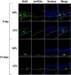
Detection of human corneal epithelial stem cells in the tissue engineered cornea. Indirect immunofluorescence analysis of BrdU (green labeling) and ΔNp63α (red labeling) to assess the presence of stem cells in the basal layer (dotted line) of the tissue-engineered epithelia produced using hCECs g...
more
Detection of human corneal epithelial stem cells in the tissue engineered cornea. Indirect immunofluorescence analysis of BrdU (green labeling) and ΔNp63α (red labeling) to assess the presence of stem cells in the basal layer (dotted line) of the tissue-engineered epithelia produced using hCECs grown with either i3T3 or iHFL as a feeder layer at 0 and 21 days following interruption of the BrdU treatment. Nuclei were counterstained with Hoechst 33,258 reagent and appear in blue. Scale bar: 20 µm.
less
Collected and cropped from Int J Mol Sci by CiteAb, provided under a CC-BY license
Image 1 of 29
In Development on 20 August 2018 by Hasenpusch-Theil, K., West, S., et al.
Fig.2.H

-
IHC-IF
-
Mus musculus (House mouse)
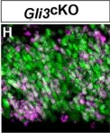
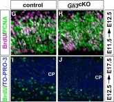
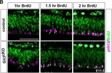
Increased proliferation and reduced cell cycle exit in Gli3cKO mutants. (A-D) Sections of E11.5 and E12.5 control and Gli3cKO embryos stained with pHH3/PCNA. (E,F) Quantification of the data presented in A-D showing increased proportions of RGCs (E11.5) and BPs (E11.5 and E12.5) undergoing mitosi...
more
Increased proliferation and reduced cell cycle exit in Gli3cKO mutants. (A-D) Sections of E11.5 and E12.5 control and Gli3cKO embryos stained with pHH3/PCNA. (E,F) Quantification of the data presented in A-D showing increased proportions of RGCs (E11.5) and BPs (E11.5 and E12.5) undergoing mitosis. (G,H) BrdU/PCNA immunohistochemistry on sections of E12.5 control and Gli3cKO embryos that were treated with BrdU 24 h earlier. (I,J) BrdU immunofluorescence staining on sections of E17.5 control (I) and Gli3cKO embryos (J) to reveal neurons born at E12.5. Only the cortical plate (CP) is shown. (K) Quantification of the data presented in G-J showing decreased cell cycle exit and neuron formation in Gli3cKO embryos. All statistical data are presented as mean±95% CI; n=6 (A,B) and n=4 (C,D,G-J); *P<0.05; Mann–Whitney test. Scale bar: in A, 25 µm for A-D,G-J.
less
Collected and cropped from Development by CiteAb, provided under a CC-BY license
Image 1 of 29
In Development on 20 August 2018 by Hasenpusch-Theil, K., West, S., et al.
Fig.2.G

-
IHC
-
Mus musculus (House mouse)
Increased proliferation and reduced cell cycle exit in Gli3cKO mutants. (A-D) Sections of E11.5 and E12.5 control and Gli3cKO embryos stained with pHH3/PCNA. (E,F) Quantification of the data presented in A-D showing increased proportions of RGCs (E11.5) and BPs (E11.5 and E12.5) undergoing mitosi...
more
Increased proliferation and reduced cell cycle exit in Gli3cKO mutants. (A-D) Sections of E11.5 and E12.5 control and Gli3cKO embryos stained with pHH3/PCNA. (E,F) Quantification of the data presented in A-D showing increased proportions of RGCs (E11.5) and BPs (E11.5 and E12.5) undergoing mitosis. (G,H) BrdU/PCNA immunohistochemistry on sections of E12.5 control and Gli3cKO embryos that were treated with BrdU 24 h earlier. (I,J) BrdU immunofluorescence staining on sections of E17.5 control (I) and Gli3cKO embryos (J) to reveal neurons born at E12.5. Only the cortical plate (CP) is shown. (K) Quantification of the data presented in G-J showing decreased cell cycle exit and neuron formation in Gli3cKO embryos. All statistical data are presented as mean±95% CI; n=6 (A,B) and n=4 (C,D,G-J); *P<0.05; Mann–Whitney test. Scale bar: in A, 25 µm for A-D,G-J.
less
Collected and cropped from Development by CiteAb, provided under a CC-BY license
Image 1 of 29
In Development on 20 August 2018 by Hasenpusch-Theil, K., West, S., et al.
Fig.4.A

-
IHC
-
Mus musculus (House mouse)
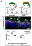
Cell cycle length of cortical progenitors is affected in Gli3cKO embryos. (A) BrdU/IdU double labelling experiments to determine total cell cycle length (TC) and S-phase duration (TS), which were calculated from counts of IdU+BrdU− and IdU+BrdU+ cells as described (Martynoga et al., 2005). E11.5 ...
more
Cell cycle length of cortical progenitors is affected in Gli3cKO embryos. (A) BrdU/IdU double labelling experiments to determine total cell cycle length (TC) and S-phase duration (TS), which were calculated from counts of IdU+BrdU− and IdU+BrdU+ cells as described (Martynoga et al., 2005). E11.5 Gli3cKO embryos show a shortening of TC and TS. The schematic illustrates the timing of IdU and BrdU injections and the progression of labelled cells in the cell cycle. (B) BrdU labelling experiments to investigate the duration of G2 (TG2). Double labelling for BrdU and pHH3 determined the proportion of BrdU+/pHH3+ cells (arrows). TG2 corresponds to the time when 50% of pHH3+ cells are BrdU+ (indicated by the dotted lines) and is not significantly altered between E11.5 control and Gli3cKO embryos. (C) Determining M-phase length (TM) using immunostaining for pHH3 and PCNA, which label mitotic and proliferating cells, respectively. The fraction of mitotic cells multiplied by total cell cycle length provides TM duration, which is not significantly altered in E11.5 Gli3cKO embryos. (D) Pie charts summarizing the length of total and individual cell cycle phases in E11.5 and E12.5 control and Gli3cKO embryos. Shorter G1 and S phases contribute to a shortening of the overall cell cycle at E11.5 whereas TC is increased at E12.5 due to longer S and M phases. All statistical data are presented as mean±95% CI; n=4; *P<0.05; Mann–Whitney test. ns, not significant. Scale bars: 50 µm.
less
Collected and cropped from Development by CiteAb, provided under a CC-BY license
Image 1 of 29
In Development on 20 August 2018 by Hasenpusch-Theil, K., West, S., et al.
Fig.4.B

-
IHC
-
Mus musculus (House mouse)
Cell cycle length of cortical progenitors is affected in Gli3cKO embryos. (A) BrdU/IdU double labelling experiments to determine total cell cycle length (TC) and S-phase duration (TS), which were calculated from counts of IdU+BrdU− and IdU+BrdU+ cells as described (Martynoga et al., 2005). E11.5 ...
more
Cell cycle length of cortical progenitors is affected in Gli3cKO embryos. (A) BrdU/IdU double labelling experiments to determine total cell cycle length (TC) and S-phase duration (TS), which were calculated from counts of IdU+BrdU− and IdU+BrdU+ cells as described (Martynoga et al., 2005). E11.5 Gli3cKO embryos show a shortening of TC and TS. The schematic illustrates the timing of IdU and BrdU injections and the progression of labelled cells in the cell cycle. (B) BrdU labelling experiments to investigate the duration of G2 (TG2). Double labelling for BrdU and pHH3 determined the proportion of BrdU+/pHH3+ cells (arrows). TG2 corresponds to the time when 50% of pHH3+ cells are BrdU+ (indicated by the dotted lines) and is not significantly altered between E11.5 control and Gli3cKO embryos. (C) Determining M-phase length (TM) using immunostaining for pHH3 and PCNA, which label mitotic and proliferating cells, respectively. The fraction of mitotic cells multiplied by total cell cycle length provides TM duration, which is not significantly altered in E11.5 Gli3cKO embryos. (D) Pie charts summarizing the length of total and individual cell cycle phases in E11.5 and E12.5 control and Gli3cKO embryos. Shorter G1 and S phases contribute to a shortening of the overall cell cycle at E11.5 whereas TC is increased at E12.5 due to longer S and M phases. All statistical data are presented as mean±95% CI; n=4; *P<0.05; Mann–Whitney test. ns, not significant. Scale bars: 50 µm.
less
Collected and cropped from Development by CiteAb, provided under a CC-BY license
Image 1 of 29
In Oncotarget on 22 September 2017 by Ghasemi, F., Black, M., et al.
Fig.4.A

-
WB
-
Homo sapiens (Human)
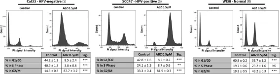
Albendazole induced cell cycle arrest at G2/M phaseCal33 (HPV-negative), SCC47 (HPV-positive) and WI38 (normal) cells were exposed to vehicle (DMSO-only) or 0.5 μM albendazole (ABZ) for 24 hours (3 replicates per treatment, ± standard error is shown). BrdU and PI staining were carried out prior t...
more
Albendazole induced cell cycle arrest at G2/M phaseCal33 (HPV-negative), SCC47 (HPV-positive) and WI38 (normal) cells were exposed to vehicle (DMSO-only) or 0.5 μM albendazole (ABZ) for 24 hours (3 replicates per treatment, ± standard error is shown). BrdU and PI staining were carried out prior to flow cytometry analysis. “(S)” marks cells lines susceptible, and “(R)” marks cell lines not susceptible to albendazole in IC50 analysis. *p < 0.05, **p < 0.01, ***p < 0.001, and NS = not significant.
less
Collected and cropped from Oncotarget by CiteAb, provided under a CC-BY license
Image 1 of 29
In Cell Death Dis on 27 July 2017 by Rohban, S., Cerutti, A., et al.
Fig.3.A

-
ICC-IF
-
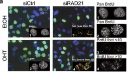
Myc-induced S-phase entry leads to replicative stress in RAD21-depleted cells. U2OS-MycER cells were transfected with siCtrl or siRAD21 and treated with either EtOH or OHT for 48 h. (a) Immunofluorescence staining of BrdU incorporation (green) at 48 h post siRNA transfection/treatment. Nuclei wer...
more
Myc-induced S-phase entry leads to replicative stress in RAD21-depleted cells. U2OS-MycER cells were transfected with siCtrl or siRAD21 and treated with either EtOH or OHT for 48 h. (a) Immunofluorescence staining of BrdU incorporation (green) at 48 h post siRNA transfection/treatment. Nuclei were stained with DAPI (in blue). Cells were pulse-labeled with BrdU for 30 min and then fixed for IF. Insets and side panels: magnification of nuclei and their classification based on BrdU incorporation. Cells with widespread nuclear incorporation of BrdU were defined as Pan-BrdU, while cells displaying only clearly identifiable focal BrdU incorporation were defined as BrdU foci. BrdU-foci-positive cells were sub-set based on the foci count, as indicated. Yellow circles highlight the BrdU-foci counted. (b) Bar graph of the percentage of BrdU-positive cells, classified as described in (a). (c) Kinetic analysis of S-phase entry of Nocodazole synchronized cells assessed by continuous BrdU incorporation. The experimental design is outlined in the upper panel. Data are presented as mean±standard deviation (Wilcoxon matched-pairs signed rank test: *P<0.05). Representative images of BrdU immunostaining at 18 h post release are shown on the right. (d) Graph of the percentage γH2AX in EdU-positive cells detected at different time points post release from a double thymidine block. Error bar represents S.D. of two independent experiments. The percentage of EdU-positive cells detected at 12 h post the first or the second thymidine release (marked by the black arrows) is indicated next to the symbols (mean value). OHT was added after the second thymidine release. (e) Quantification of RPA foci determined by immunofluorescence (48 h post transfection). Left: representative images of foci in RPA-positive nuclei; right: dot-plot, number of cells is indicated within parentheses. Significant differences are indicated by the asterisks (t-test: *P<0.05). Effect size (Cohen’s d): +OHT (siCtrl versus siRAD21)=0.36
less
Collected and cropped from Cell Death Dis by CiteAb, provided under a CC-BY license
Image 1 of 29
In Cell Death Dis on 9 March 2017 by Deshmukh, J., Pofahl, R., et al.
Fig.8.C

-
IHC
-
Mus musculus (House mouse)
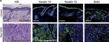
Epidermis-specific deletion of Rac1 facilitates development of SCC upon long-term UV-irradiation. (a and b) Representative photographs of the mice with hyperkeratotic papules in healed skin erosions of Rac1-EKO mice. The image in b is a close-up of the lesion seen in the Rac1-EKO mouse in (a). (c...
more
Epidermis-specific deletion of Rac1 facilitates development of SCC upon long-term UV-irradiation. (a and b) Representative photographs of the mice with hyperkeratotic papules in healed skin erosions of Rac1-EKO mice. The image in b is a close-up of the lesion seen in the Rac1-EKO mouse in (a). (c) H/E staining and immunostainings against keratin 14, keratin 10, and BrdU (green) of skin and tumor samples of controls and Rac1-EKO mice. Nuclei are stained in blue. Scale bar=100 μm. (b): Graph shows the results of the histological analysis of skin samples or tumors from control and Rac1-EKO mice. Mice without tumors are shown in blue and mice with SCCs in gray. Absolute numbers of mice are given within the bars
less
Collected and cropped from Cell Death Dis by CiteAb, provided under a CC-BY license
Image 1 of 29
In PLoS Pathog on 1 March 2017 by Xu, P., Zhou, Z., et al.
Fig.7.A

-
FC/FACS
-
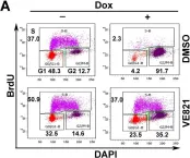
Homo sapiens (Human)
Inhibiting ATR phosphorylation abolishes NS1-induced G2-phase arrest in UT7/Epo-S1 cells.(A) Cell cycle analysis. NS1-S1 cells were treated with the ATR-specific inhibitor VE821 at 3 h prior to Dox treatment. At 72 h post-treatment, the cells were then collected and co-stained with an anti-BrdU a...
more
Inhibiting ATR phosphorylation abolishes NS1-induced G2-phase arrest in UT7/Epo-S1 cells.(A) Cell cycle analysis. NS1-S1 cells were treated with the ATR-specific inhibitor VE821 at 3 h prior to Dox treatment. At 72 h post-treatment, the cells were then collected and co-stained with an anti-BrdU antibody and DAPI prior for flow cytometry. DMSO-treated NS1-S1 cells were used as a control. (B) Statistical analyses. The percentage of the cells at each stage of the cell cycle is depicted in color. Numbers shown are the percentages at G2-phase and are statistically compared within cell groups treated with or without Dox induction as indicated. **P<0.01, and N.S. represents no significance. (C) ATR inhibition. After treatment with DMSO or VE821, cells were collected, and expression of ATR(pT1989) was examined by Western blotting. (D) Activation of the ATR-CDC25C-CDK1 pathway. NS1-S1 cells were either transduced with lentivirus harboring scramble or ATR-specific shRNA for 48 h, or treated with DMSO and VE821, and then treated with Dox for 72 h. The cells were then collected, lysed, and immunoblotted with the indicated antibodies. (E&F) Quantification. The detected bands of CDC25C(pS216) and CDK1(pY15) shown in panel D were quantified, and the results are expressed as the mean ± standard deviation of at least three independent experiments. Statistical analysis was performed in paired groups as indicated. **P<0.01, and *P<0.05.
less
Collected and cropped from PLoS Pathog by CiteAb, provided under a CC-BY license
Image 1 of 29
In Sci Rep on 23 August 2016 by Halin Bergström, S., Hägglöf, C., et al.
Fig.3.B

-
IHC
-
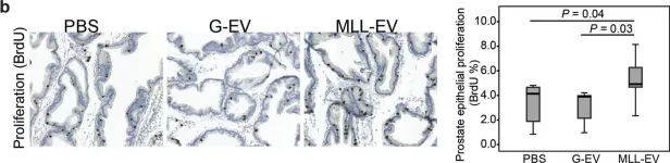
Rattus norvegicus (Rat)
Immunostaining in rat prostates stimulated with tumor-derived extracellular vesicles (EVs).Rat ventral prostates were injected with PBS (n = 6), G-EVs (n = 6), or MLL-EVs (n = 6) and the prostate tissue was analyzed after 72 hours. Sections were immunostained and quantified for (a) macrophages (C...
more
Immunostaining in rat prostates stimulated with tumor-derived extracellular vesicles (EVs).Rat ventral prostates were injected with PBS (n = 6), G-EVs (n = 6), or MLL-EVs (n = 6) and the prostate tissue was analyzed after 72 hours. Sections were immunostained and quantified for (a) macrophages (CD68) and (b) epithelial proliferation (BrdU). Sections show representative staining (brown) in each group (original magnification x200). Each group was illustrated with box plots.
less
Collected and cropped from Sci Rep by CiteAb, provided under a CC-BY license
Image 1 of 29
In Open Biol on 1 January 2016 by Ligasová, A., Liboska, R., et al.
Fig.1.A

-
ICC-IF
-
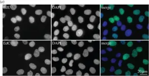
Homo sapiens (Human)
The microscopy analysis of EdC conversion to EdU using an antibody reaction. (a) Fluorescence detection of EdU by means of an anti-bromodeoxyuridine antibody (clone B44). HeLa cells were incubated with either 10 µM EdU or 10 µM EdC. Then, the detection of EdU (in green) and DNA using DAPI (in blu...
more
The microscopy analysis of EdC conversion to EdU using an antibody reaction. (a) Fluorescence detection of EdU by means of an anti-bromodeoxyuridine antibody (clone B44). HeLa cells were incubated with either 10 µM EdU or 10 µM EdC. Then, the detection of EdU (in green) and DNA using DAPI (in blue) was performed. (b) The analysis of the reactivity of anti-bromodeoxyuridine antibody (clone B44) using EdU with biotin at the 5′ end and EdC with biotin at the 3′ or 5′ end. The data were normalized to percentage of the signal provided by EdU (equal to 100%). The data are presented as mean ± s.e.m.
less
Collected and cropped from Open Biol by CiteAb, provided under a CC-BY license
Image 1 of 29
In Open Biol on 1 January 2016 by Ligasová, A., Liboska, R., et al.
Fig.6.A

-
ICC-IF
-
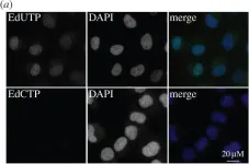
Homo sapiens (Human)
Run-on replication assay and hypotonic introduction of EdUTP and EdCTP, EdCDP and EdCMP. (a) The detection of EdU and EdC using Alexa Fluor 488 azide in permeabilized HeLa cells (in green). The nuclear DNA was stained by DAPI (in blue). (b) The average signal in cell nuclei after detection of EdU...
more
Run-on replication assay and hypotonic introduction of EdUTP and EdCTP, EdCDP and EdCMP. (a) The detection of EdU and EdC using Alexa Fluor 488 azide in permeabilized HeLa cells (in green). The nuclear DNA was stained by DAPI (in blue). (b) The average signal in cell nuclei after detection of EdU and EdC by Alexa Fluor 488 azide or EdU by the anti-bromodeoxyuridine antibody clone B44 in HeLa cells after the hypotonic introduction of EdUTP and EdCTP followed by a 30-min incubation in medium. The data are normalized to percentage of the signal of EdUTP-treated cells (equal to 100%). The data are presented as mean ± s.e.m. (c) The average signal in cell nuclei after detection of EdU and EdC by Alexa Fluor 488 azide or EdU by the anti-bromodeoxyuridine antibody clone B44 in HeLa cells after the hypotonic introduction of EdCMP, EdCDP and EdCTP followed by a 30-min incubation in medium. The data are normalized to percentage of the signal of EdCMP-treated cells (equal to 100%). The data are presented as mean ± s.e.m. (d) The average signal in cell nuclei after detection of EdU and EdC by Alexa Fluor 488 azide in HeLa cells after the hypotonic introduction of EdUTP or EdCTP with the concurrent introduction of dTTP. The data are normalized to percentage of the signal of EdUTP- or EdCTP-treated cells (equal to 100%). The data are presented as mean ± s.e.m.
less
Collected and cropped from Open Biol by CiteAb, provided under a CC-BY license
Image 1 of 29
In Aging Cell on 1 February 2015 by Boquoi, A., Arora, S., et al.
Fig.2.A

-
IHC
-
Mus musculus (House mouse)
Induced p16 inhibits proliferation of intestinal stem cells. (a) CMV-rtTA:TetO-p16:Lgr5-lacZ mice were treated with Dox for 1 week and injected with BrdU. LacZ was detected by activity stain (left, aqua), p16 by IHC (left, brown), and BrdU in the same section by IF (right, red). Note paucity of B...
more
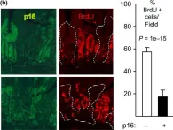
Induced p16 inhibits proliferation of intestinal stem cells. (a) CMV-rtTA:TetO-p16:Lgr5-lacZ mice were treated with Dox for 1 week and injected with BrdU. LacZ was detected by activity stain (left, aqua), p16 by IHC (left, brown), and BrdU in the same section by IF (right, red). Note paucity of BrdU staining in the p16+ Lgr5-lacZ+ cells (left 3 crypts). Across 8 20× fields, p16+ Lgr5+ cells (N = 331) showed no BrdU staining vs. 15% of the p16- Lgr5+ cells (N = 461, P = 0.02) (b) In the same mice, p16 and BrdU were detected by IHC in serial 4- to 5-micrometer sections and the % BrdU+ cells were scored in Lgr5+ and transit-amplifying cells, respectively. P values by two-sided t-tests. The one-sided t-test for transit cells = 0.03. N = 3 mice; 5803 cells counted. (c) Reduced p16 expression in small intestines of mice with continuous p16 induction. CMV-rtTA:TetO-p16:Lgr5-lacZ mice were treated with Dox near 2 mos of age for 1 or 4 weeks. IHC for p16 (brown) and activity assay for lacZ (aqua). Fields 10x. N = 3 mice per time point. Note the gradual loss of p16 expression, with preferential retention at the crypt base (P = 0.02 vs. transit-amplifying zones, P = 0.007 vs. villi). An atrophic, p16+ crypt is marked. (d, e) Partial costaining for p16 and lysozyme in crypt base cells, detected by confocal co-IF. Mice treated with Dox d20-40 were stained for p16 (green), lysozyme (red), and DNA (DAPI, blue; top). Green arrows (d) mark a p16+ cell that stains weakly for lysozyme, reflecting partial paneth cell differentiation. White arrows (e) identify a p16+ lysozyme- cell that is surrounded by lysozyme+ cells and, hence, is likely a stem cell.
less
Collected and cropped from Aging Cell by CiteAb, provided under a CC-BY license
Image 1 of 29
In Aging Cell on 1 February 2015 by Boquoi, A., Arora, S., et al.
Fig.1.B

-
IHC
-
Mus musculus (House mouse)
p16 induction inhibits intestinal epithelial cell proliferation. (a) Mice of different genotypes were or were not treated with Dox for 1 week, as designated. IHC for exogenous p16 (brown, 10× fields). Note strong mosaic p16 induction in the presence of both transgenes and Dox. (b) CMV-rtTA:TetO-p...
more
p16 induction inhibits intestinal epithelial cell proliferation. (a) Mice of different genotypes were or were not treated with Dox for 1 week, as designated. IHC for exogenous p16 (brown, 10× fields). Note strong mosaic p16 induction in the presence of both transgenes and Dox. (b) CMV-rtTA:TetO-p16-1 mice treated with Dox for 1 week and injected with BrdU. Panels (left): Two 20× fields (above, below) with co-IF for p16 (left, green; right, dashed lines) and BrdU (right, red). Graph (right): Among 1122 crypt cells scored, 19% of p16+ cells were BrdU+ vs. 58% of p16- cells. Modest BrdU signal was visible in the green filter.
less
Collected and cropped from Aging Cell by CiteAb, provided under a CC-BY license
Image 1 of 29
In Regeneration (Oxf) on 1 February 2014 by Wu, P., Alibardi, L., et al.
Fig.1.K,L,M

-
IHC-IF
-
Lizard
Scale neogenesis on regenerating tail of A. carolinensis. (A)−(D) Gross morphology of A. carolinensis tails at 12, 19, 26 and 33 days of regeneration. (A) Dark blastema (arrow); (B) elongating cone; (C1) regenerating tail showing the beginning of scale formation; (D1) regenerated tail with scalin...
more
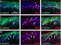
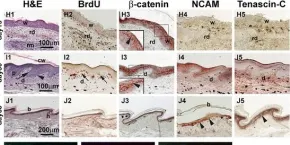
Scale neogenesis on regenerating tail of A. carolinensis. (A)−(D) Gross morphology of A. carolinensis tails at 12, 19, 26 and 33 days of regeneration. (A) Dark blastema (arrow); (B) elongating cone; (C1) regenerating tail showing the beginning of scale formation; (D1) regenerated tail with scaling in proximal regions; (C2), (D2) enlargements from the proximal part of (C1) and (D1), respectively. (E)−(J1) Skin histology with hematoxylin and eosin (H&E) stained sections: (E) blastema; (F) elongating cone (the arrowhead indicates the wound epidermis); (G) scaled tail (the arrowhead points to regenerated scales); (H1) thick wound epidermis before scaling begins; (I1) epidermal pegs in scaling epidermis; (J1) regenerated scales. (H2)−(J5) Immunocytochemistry for specific markers. (H2)−(J2) BrdU staining. (H3)−(J3) β‐catenin staining. Inserts in H3 and I3 show enlargements of the indicated areas (arrowheads indicate membranous immunostaining of keratinocytes). (H4)−(J4) NCAM staining (arrowheads indicate more intense staining). (H5)−(J5) Tenascin‐C staining (the arrowhead indicates a more intensely labeled region). (K)−(M) Confocal immunocytochemistry of day 30 regenerating skin. Left panel, β‐catenin staining. Middle panel, BrdU staining. Right panel, combined. (K) Double‐labeled nuclei (arrow) in the undulated wound epidermis with diffuse β‐catenin labeling in suprabasal layers. (L) Early peg showing double‐labeled nuclei (arrow) and β‐catenin in suprabasal keratinocytes. (M) Elongated peg with double‐labeled nuclei (arrow) and BrdU single‐labeled nuclei (arrowhead). Double arrowheads in (L) and (M) indicate the β‐catenin nuclear positive cells in the mesenchyme. b, beta‐layer; bl, blastema; ca, regenerating cartilage; d, dermis; e, ependyma; h, hinge; l, lacunar epithelium; p, epidermal peg; nt, normal tail; rd, regenerated dermis; rm, regenerated muscle; rs, regenerated scale; sb, suprabasal layer; se, scaling epidermis; w, wound epidermis.
less
Collected and cropped from Regeneration (Oxf) by CiteAb, provided under a CC-BY license
Image 1 of 29
In Regeneration (Oxf) on 1 February 2014 by Wu, P., Alibardi, L., et al.
Fig.1.H,I,J

-
IHC
-
Lizard
Scale neogenesis on regenerating tail of A. carolinensis. (A)−(D) Gross morphology of A. carolinensis tails at 12, 19, 26 and 33 days of regeneration. (A) Dark blastema (arrow); (B) elongating cone; (C1) regenerating tail showing the beginning of scale formation; (D1) regenerated tail with scalin...
more
Scale neogenesis on regenerating tail of A. carolinensis. (A)−(D) Gross morphology of A. carolinensis tails at 12, 19, 26 and 33 days of regeneration. (A) Dark blastema (arrow); (B) elongating cone; (C1) regenerating tail showing the beginning of scale formation; (D1) regenerated tail with scaling in proximal regions; (C2), (D2) enlargements from the proximal part of (C1) and (D1), respectively. (E)−(J1) Skin histology with hematoxylin and eosin (H&E) stained sections: (E) blastema; (F) elongating cone (the arrowhead indicates the wound epidermis); (G) scaled tail (the arrowhead points to regenerated scales); (H1) thick wound epidermis before scaling begins; (I1) epidermal pegs in scaling epidermis; (J1) regenerated scales. (H2)−(J5) Immunocytochemistry for specific markers. (H2)−(J2) BrdU staining. (H3)−(J3) β‐catenin staining. Inserts in H3 and I3 show enlargements of the indicated areas (arrowheads indicate membranous immunostaining of keratinocytes). (H4)−(J4) NCAM staining (arrowheads indicate more intense staining). (H5)−(J5) Tenascin‐C staining (the arrowhead indicates a more intensely labeled region). (K)−(M) Confocal immunocytochemistry of day 30 regenerating skin. Left panel, β‐catenin staining. Middle panel, BrdU staining. Right panel, combined. (K) Double‐labeled nuclei (arrow) in the undulated wound epidermis with diffuse β‐catenin labeling in suprabasal layers. (L) Early peg showing double‐labeled nuclei (arrow) and β‐catenin in suprabasal keratinocytes. (M) Elongated peg with double‐labeled nuclei (arrow) and BrdU single‐labeled nuclei (arrowhead). Double arrowheads in (L) and (M) indicate the β‐catenin nuclear positive cells in the mesenchyme. b, beta‐layer; bl, blastema; ca, regenerating cartilage; d, dermis; e, ependyma; h, hinge; l, lacunar epithelium; p, epidermal peg; nt, normal tail; rd, regenerated dermis; rm, regenerated muscle; rs, regenerated scale; sb, suprabasal layer; se, scaling epidermis; w, wound epidermis.
less
Collected and cropped from Regeneration (Oxf) by CiteAb, provided under a CC-BY license
Image 1 of 29
In PLoS One on 12 April 2012 by Reich, J. & Papoulas, O.
Fig.5.D

-
ICC-IF
-
Mus musculus (House mouse)
Loss of Capr alters cell cycle dynamics but not proliferation rates in the FSC lineage.A-C) Fixed Df(3L)Cat/+ (control), or Df(3L)Cat/Capr2 (Capr-) germaria were stained with antibodies to phospho-histone H3 (red), and FasIII (green), and with TO-PRO-3 iodide (blue). A) Quantification of the % of...
more
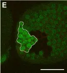
Loss of Capr alters cell cycle dynamics but not proliferation rates in the FSC lineage.A-C) Fixed Df(3L)Cat/+ (control), or Df(3L)Cat/Capr2 (Capr-) germaria were stained with antibodies to phospho-histone H3 (red), and FasIII (green), and with TO-PRO-3 iodide (blue). A) Quantification of the % of germaria scored that showed any phospho-histone H3-positive staining in cells of the FSC lineage. The number of germaria analysed was 81 control, 82 Capr-. B-C) Examples of stained germaria of the indicated genotypes. Size bar is 30 microns. D) Quantification of the % of pulse labeled germaria scored that incorporated BrdU in cells of the FSC lineage. The difference between Df(3L)Cat/+ (control), or Df(3L)Cat/Capr2 (Capr-) was not significant (P = 0.09). The number of germaria analysed was 80 control, 86 Capr-. E) Tangential section of a fixed stage-10 egg chamber stained for GFP (green) and FasIII (red). The Capr- follicle cell clone (no GFP staining) and its adjacent wild-type twin-spot (bright green) are of similar size. Size bar is 60 microns.
less
Collected and cropped from PLoS One by CiteAb, provided under a CC-BY license
Image 1 of 29