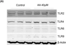
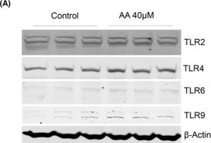
Aristolochic acids (AAs) are extracted from certain plants as folk remedies for centuries until their nephrotoxicity and carcinogenicity were recognized. Aristolochic acid I (AAI) is one of the main pathogenic compounds, and it has nephrotoxic, carcinogenic and mutagenic effects. Previous studies have shown that AAI acts mainly on proximal renal tubular epithelial cells; however, the mechanisms of AAI-induced proximal tubule cell damage are still not fully characterized. We exposed human kidney proximal tubule cells (PTCs; HK2 cell line) to AAI in vitro at different time/dose conditions and assessed cell proliferation, reactive oxygen species (ROS) generation, nitric oxide (NO) production, m-RNA/ protein expressions and mitochondrial dysfunction. AAI exposure decreased proliferation and increased apoptosis, ROS generation / NO production in PTCs significantly at 24 h. Gene/ protein expression studies demonstrated activation of innate immunity (TLRs 2, 3, 4 and 9, HMGB1), inflammatory (IL6, TNFA, IL1B, IL18, TGFB and NLRP3) and kidney injury (LCN2) markers. AAI also induced epithelial-mesenchymal transition (EMT) and mitochondrial dysfunction in HK2 cells. TLR9 knock-down and ROS inhibition were able to ameliorate the toxic effect of AAI. In conclusion, AAI treatment caused injury to PTCs through ROS-HMGB1/mitochondrial DNA (mt DNA)-mediated activation of TLRs and inflammatory response.
© 2022 The Authors. Journal of Cellular and Molecular Medicine published by Foundation for Cellular and Molecular Medicine and John Wiley & Sons Ltd.
Product Citations: 2
Applications
Reactivity
Research Area
In Journal of Cellular and Molecular Medicine on 1 August 2022 by Upadhyay, R. & Batuman, V.
-
WB
-
Mus musculus (House mouse)
-
Biochemistry and Molecular biology
-
Genetics
Free light chains injure proximal tubule cells through the STAT1/HMGB1/TLR axis.
In JCI Insight on 23 July 2020 by Upadhyay, R., Ying, W. Z., et al.
Free light chains (FLCs) induce inflammatory pathways in proximal tubule cells (PTCs). The role of TLRs in these responses is unknown. Here we present findings on the role of TLRs in FLC-induced PTC injury. We exposed human kidney PTC cultures to κ and λ FLCs and used cell supernatants and pellets for ELISA and gene expression studies. We also analyzed tissues from Stat1-/- and littermate control mice treated with daily i.p. injections of a κ FLC for 10 days. FLCs increased the expression of TLR2, TLR4, and TLR6 via HMGB1, a damage-associated molecular pattern. Countering TLR2, TLR4, and TLR6 through GIT-27 or specific TLR siRNAs reduced downstream cytokine responses. Blocking HMGB1 through siRNA or pharmacologic inhibition, or via STAT1 inhibition, reduced FLC-induced TLR2, TLR4, and TLR6 expression. Blocking endocytosis of FLCs through silencing of megalin/cubilin, with bafilomycin A1 or hypertonic sucrose, attenuated FLC-induced cytokine responses in PTCs. IHC showed decreased TLR4 and TLR6 expression in kidney sections from Stat1-/- mice compared with their littermate controls. PTCs exposed to FLCs released HMGB1, which induced expression of TLR2, TLR4, and TLR6 and downstream inflammation. Blocking FLCs' endocytosis, Stat1 knockdown, HMGB1 inhibition, and TLR knockdown each rescued PTCs from FLC-induced injury.
-
WB
In J Cell Mol Med on 1 August 2022 by Upadhyay, R. & Batuman, V.
Fig.3.A
-
WB
-
Collected and cropped from Journal of Cellular and Molecular Medicine by CiteAb, provided under a CC-BY license
Image 1 of 1