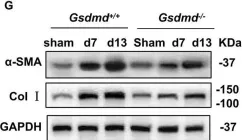
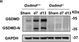
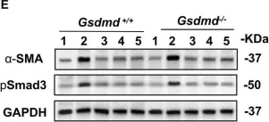


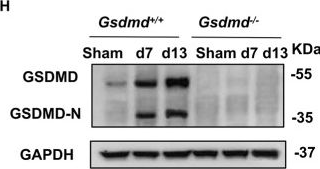
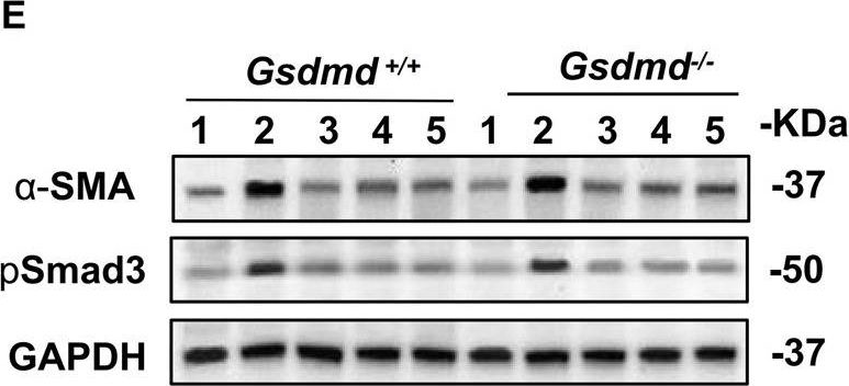
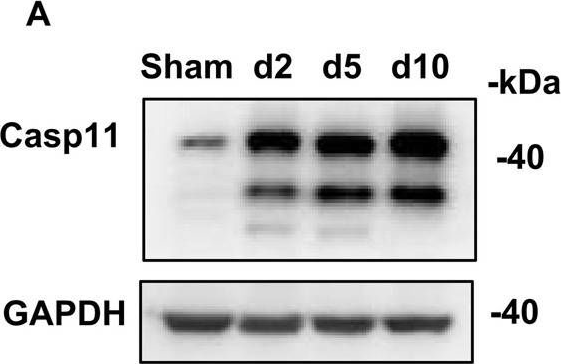
Renal fibrosis is a common consequence of various progressive nephropathies, including obstructive nephropathy, and ultimately leads to kidney failure. Infiltration of inflammatory cells is a prominent feature of renal injury after draining blockages from the kidney, and correlates closely with the development of renal fibrosis. However, the underlying molecular mechanism behind the promotion of renal fibrosis by inflammatory cells remains unclear. Herein, we showed that unilateral ureteral obstruction (UUO) induced Gasdermin D (GSDMD) activation in neutrophils, abundant neutrophil extracellular traps (NETs) formation and macrophage-to-myofibroblast transition (MMT) characterized by α-smooth muscle actin (α-SMA) expression in macrophages. Gsdmd deletion significantly reduced infiltration of inflammatory cells in the kidneys and inhibited NETs formation, MMT and thereby renal fibrosis. Chimera studies confirmed that Gsdmd deletion in bone marrow-derived cells, instead of renal parenchymal cells, provided protection against renal fibrosis. Further, specific deletion of Gsdmd in neutrophils instead of macrophages protected the kidney from undergoing fibrosis after UUO. Single-cell RNA sequencing identified robust crosstalk between neutrophils and macrophages. In vitro, GSDMD-dependent NETs triggered p65 translocation to the nucleus, which boosted the production of inflammatory cytokines and α-SMA expression in macrophages by activating TGF-β1/Smad pathway. In addition, we demonstrated that caspase-11, that could cleave GSDMD, was required for NETs formation and renal fibrosis after UUO. Collectively, our findings demonstrate that caspase-11/GSDMD-dependent NETs promote renal fibrosis by facilitating inflammation and MMT, therefore highlighting the role and mechanisms of NETs in renal fibrosis.
© 2022. The Author(s).
Product Citations: 10
Applications
Reactivity
Research Area
In Cell Death & Disease on 8 August 2022 by Wang, Y., Li, Y., et al.
-
WB
-
Mus musculus (House mouse)
-
Cell Biology
-
Immunology and Microbiology
GSDME-mediated pyroptosis promotes inflammation and fibrosis in obstructive nephropathy.
In Cell Death and Differentiation on 1 August 2021 by Li, Y., Yuan, Y., et al.
Renal tubular cell (RTC) death and inflammation contribute to the progression of obstructive nephropathy, but its underlying mechanisms have not been fully elucidated. Here, we showed that Gasdermin E (GSDME) expression level and GSDME-N domain generation determined the RTC fate response to TNFα under the condition of oxygen-glucose-serum deprivation. Deletion of Caspase-3 (Casp3) or Gsdme alleviated renal tubule damage and inflammation and finally prevented the development of hydronephrosis and kidney fibrosis after ureteral obstruction. Using bone marrow transplantation and cell type-specific Casp3 knockout mice, we demonstrated that Casp3/GSDME-mediated pyroptosis in renal parenchymal cells, but not in hematopoietic cells, played predominant roles in this process. We further showed that HMGB1 released from pyroptotic RTCs amplified inflammatory responses, which critically contributed to renal fibrogenesis. Specific deletion of Hmgb1 in RTCs alleviated caspase11 and IL-1β activation in macrophages. Collectively, our results uncovered that TNFα/Casp3/GSDME-mediated pyroptosis is responsible for the initiation of ureteral obstruction-induced renal tubule injury, which subsequentially contributes to the late-stage progression of hydronephrosis, inflammation, and fibrosis. This novel mechanism will provide valuable therapeutic insights for the treatment of obstructive nephropathy.
© 2021. The Author(s), under exclusive licence to ADMC Associazione Differenziamento e Morte Cellulare.
-
WB
-
Cell Biology
-
Immunology and Microbiology
In Journal of Virology on 9 December 2020 by Bamia, A., Marcato, V., et al.
Rift Valley fever virus (RVFV) is a highly pathogenic zoonotic arbovirus endemic in many African countries and the Arabian Peninsula. Animal infections cause high rates of mortality and abortion among sheep, goats, and cattle. In humans, an estimated 1 to 2% of RVFV infections result in severe disease (encephalitis, hepatitis, or retinitis) with a high rate of lethality when associated with hemorrhagic fever. The RVFV NSs protein, which is the main virulence factor, counteracts the host innate antiviral response to favor viral replication and spread. However, the mechanisms underlying RVFV-induced cytopathic effects and the role of NSs in these alterations remain for the most part unknown. In this work, we have analyzed the effects of NSs expression on the actin cytoskeleton while conducting infections with the NSs-expressing virulent (ZH548) and attenuated (MP12) strains of RVFV and the non-NSs-expressing avirulent (ZH548ΔNSs) strain, as well as after the ectopic expression of NSs. In macrophages, fibroblasts, and hepatocytes, NSs expression prevented the upregulation of Abl2 (a major regulator of the actin cytoskeleton) expression otherwise induced by avirulent infections and identified here as part of the antiviral response. The presence of NSs was also linked to an increased mobility of ZH548-infected cells compared to ZH548ΔNSs-infected fibroblasts and to strong changes in cell morphology in nonmigrating hepatocytes, with reduction of lamellipodia, cell spreading, and dissolution of adherens junctions reminiscent of the ZH548-induced cytopathic effects observed in vivo Finally, we show evidence of the presence of NSs within long actin-rich structures associated with NSs dissemination from NSs-expressing toward non-NSs-expressing cells.IMPORTANCE Rift Valley fever virus (RVFV) is a dangerous human and animal pathogen that was ranked by the World Health Organization in 2018 as among the eight pathogens of most concern for being likely to cause wide epidemics in the near future and for which there are no, or insufficient, countermeasures. The focus of this work is to address the question of the mechanisms underlying RVFV-induced cytopathic effects that participate in RVFV pathogenicity. We demonstrate here that RVFV targets cell adhesion and the actin cytoskeleton at the transcriptional and cellular level, affecting cell mobility and inducing cell shape collapse, along with distortion of cell-cell adhesion. All these effects may participate in RVFV-induced pathogenicity, facilitate virulent RVFV dissemination, and thus constitute interesting potential targets for future development of antiviral therapeutic strategies that, in the case of RVFV, as with several other emerging arboviruses, are presently lacking.
Copyright © 2020 American Society for Microbiology.
-
Cell Biology
-
Immunology and Microbiology
RIPK3 collaborates with GSDMD to drive tissue injury in lethal polymicrobial sepsis.
In Cell Death and Differentiation on 1 September 2020 by Chen, H., Li, Y., et al.
Sepsis is a systemic inflammatory disease causing life-threatening multi-organ dysfunction. Accumulating evidences suggest that two forms of programmed necrosis, necroptosis and pyroptosis triggered by the pathogen component lipopolysaccharide (LPS) and inflammatory cytokines, play important roles in the development of bacterial sepsis-induced shock and tissue injury. Sepsis-induced shock and tissue injury required receptor-interacting protein kinase-3 (RIPK3) and mixed lineage kinase domain-like protein (MLKL) phosphorylation, caspase11 activation and gasdermin D (GSDMD) cleavage. However, the synergistic effect of necroptosis and pyroptosis in the pathological progress of sepsis remains elusive. In this study, we found that blockage of both necroptosis and pyroptosis (double deletion of Ripk3/Gsdmd or Mlkl/Gsdmd) resulted in accumulative protection against septic shock, systemic blood clotting and multi-organ injury in mice. Bone marrow transplantation confirmed that necroptosis and pyroptosis in both myeloid and nonmyeloid cells are indispensable in the progression of sepsis-induced multi-organ injury. Both RIPK3 and GSDMD signaling collaborated to amplify necroinflammation and tissue factor release in macrophages and endothelial cells, which led to tissue injury. Furthermore, cell death induced by inflammatory cytokines and high-mobility group box 1 could be prevented by double ablation of Ripk3/Gsdmd or Mlkl/Gsdmd, suggesting that a positive feedback loop interconnecting RIPK3/MLKL and GSDMD machinery and inflammation facilitated sepsis progression. Collectively, our findings demonstrated that RIPK3-mediated necroptosis and GSDMD-mediated pyroptosis collaborated to amply inflammatory signaling and enhance tissue injury in the process of sepsis, which may shed new light on two potential targets of combined therapeutic interventions for this highly lethal disorder.
-
WB
-
Cell Biology
In Frontiers in Cell and Developmental Biology on 28 August 2020 by Assar, E. A. & Tumbarello, D. A.
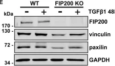
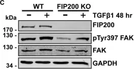
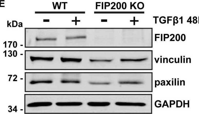
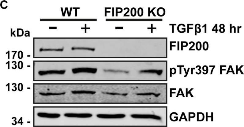
Autophagy is an essential catabolic intracellular pathway that maintains homeostasis by degrading long-lived proteins, damaged organelles, and provides an energy source during nutrient starvation. It is now understood that autophagy has discrete functions as a selective lysosomal degradation pathway targeting large cytosolic structural and signaling complexes to influence cell motility and adhesion. We provide evidence suggesting the primary autophagy regulators Atg5 and FIP200 both play a role in cell motility and extracellular matrix adhesion. However, their loss of function has a differential impact on focal adhesion composition and organization, as well as signaling in response to fibronectin induced cell spreading. This differential impact on focal adhesions is illustrated by smaller focal adhesion complexes and a decrease in FAK, paxillin, and vinculin expression associated with FIP200 loss of function. In contrast, Atg5 loss of function results in production of large and stable focal adhesions, characterized by their retention of phosphorylated FAK and Src, which correlates with increased vinculin and FAK protein expression. Importantly, autophagy is upregulated during processes associated with focal adhesion reorganization and their exhibits colocalization of autophagosomes with focal adhesion cargo. Interestingly, FIP200 localizes to vinculin-rich focal adhesions and its loss negatively regulates FAK phosphorylation. These data collectively suggest FIP200 and Atg5 may have both autophagy-dependent and -independent functions that provide distinct mechanisms and impacts on focal adhesion dynamics associated with cell motility.
Copyright © 2020 Assar and Tumbarello.
-
WB
-
Homo sapiens (Human)
-
Cell Biology
In Cell Death Dis on 8 August 2022 by Wang, Y., Li, Y., et al.
Fig.1.G

-
WB
-
Mus musculus (House mouse)
Collected and cropped from Cell Death & Disease by CiteAb, provided under a CC-BY license
Image 1 of 7
In Cell Death Dis on 8 August 2022 by Wang, Y., Li, Y., et al.
Fig.1.H
-
WB
-
Mus musculus (House mouse)
Collected and cropped from Cell Death & Disease by CiteAb, provided under a CC-BY license
Image 1 of 7
In Cell Death Dis on 8 August 2022 by Wang, Y., Li, Y., et al.
Fig.7.E
-
WB
-
Mus musculus (House mouse)
Collected and cropped from Cell Death & Disease by CiteAb, provided under a CC-BY license
Image 1 of 7
In Cell Death Dis on 8 August 2022 by Wang, Y., Li, Y., et al.
Fig.8.A
-
WB
-
Mus musculus (House mouse)
Collected and cropped from Cell Death & Disease by CiteAb, provided under a CC-BY license
Image 1 of 7
In Front Cell Dev Biol on 28 August 2020 by Assar, E. A. & Tumbarello, D. A.
Fig.5.E
-
WB
-
Homo sapiens (Human)
Collected and cropped from Frontiers in Cell and Developmental Biology by CiteAb, provided under a CC-BY license
Image 1 of 7
In Front Cell Dev Biol on 28 August 2020 by Assar, E. A. & Tumbarello, D. A.
Fig.5.C
-
WB
-
Homo sapiens (Human)
Collected and cropped from Frontiers in Cell and Developmental Biology by CiteAb, provided under a CC-BY license
Image 1 of 7
In Cell Death Dis on 29 August 2018 by Chen, H., Fang, Y., et al.
Fig.1.A
-
WB
-
Collected and cropped from Cell Death & Disease by CiteAb, provided under a CC-BY license
Image 1 of 7