Extracellular vesicles (EVs) have crucial roles in hemostasis and coagulation. They sustain coagulation by exposing phosphatidylserine and initiate clotting by surface expression of tissue factor (TF) under inflammatory conditions. As their relevance as biomarkers of coagulopathy is increasingly recognized, there is a need for the sensitive and reliable detection of TF+ EVs, but their flow cytometric analysis is challenging and has yielded controversial findings for TF expression on EVs in the vascular system. We investigated the effect of different fluorochrome-to-protein (F/P) ratios of anti-TF-fluorochrome conjugates on the flow cytometric detection of TF+ EVs from activated monocytes, mesenchymal stem cells (MSCs), and in COVID-19 plasma. Using a FITC-labeled anti-TF antibody (clone VD8), we show that the percentage of TF+ EVs declined with decreasing F/P ratios. TF was detected on 7.6%, 5.4%, and 1.1% of all EVs derived from activated monocytes at F/P ratios of 7.7:1, 6.6:1, and 5.2:1. A similar decline was observed for EVs from MSCs and for EVs in plasma, whereas the detection of TF on cells remained unaffected by different F/P ratios. We provide clear evidence that next to the antibody clone, the F/P ratio affects the flow cytometric detection of TF+ EVs and should be carefully controlled.
© 2024. The Author(s).
Product Citations: 16
In Scientific Reports on 17 March 2024 by Weiss, R., Mostageer, M., et al.
Tissue Factor and COVID-19 Associated Thrombosis.
In Arteriosclerosis, Thrombosis, and Vascular Biology on 1 March 2024 by Mackman, N.
Microbial infections activate the innate and adaptive immune systems.1 Pathogen-associated molecular patterns produced by microbes, such as double-stranded RNA, are detected by PRRs (pattern-recognition receptors), such as toll-like receptor 3, and this leads to the expression of interferons and cytokines.1,2.
-
Cardiovascular biology
-
COVID-19
Reduced monocyte proportions and responsiveness in convalescent COVID-19 patients.
In Frontiers in Immunology on 22 January 2024 by Ravkov, E. V., Williams, E. S. C. P., et al.
The clinical manifestations of acute severe acute respiratory syndrome coronavirus type 2 (SARS-CoV-2) infection and coronavirus disease 2019 (COVID-19) suggest a dysregulation of the host immune response that leads to inflammation, thrombosis, and organ dysfunction. It is less clear whether these dysregulated processes persist during the convalescent phase of disease or during long COVID. We sought to examine the effects of SARS-CoV-2 infection on the proportions of classical, intermediate, and nonclassical monocytes, their activation status, and their functional properties in convalescent COVID-19 patients.
Peripheral blood mononuclear cells (PBMCs) from convalescent COVID-19 patients and uninfected controls were analyzed by multiparameter flow cytometry to determine relative percentages of total monocytes and monocyte subsets. The expression of activation markers and proinflammatory cytokines in response to LPS treatment were measured by flow cytometry and ELISA, respectively.
We found that the percentage of total monocytes was decreased in convalescent COVID-19 patients compared to uninfected controls. This was due to decreased intermediate and non-classical monocytes. Classical monocytes from convalescent COVID-19 patients demonstrated a decrease in activation markers, such as CD56, in response to stimulation with bacterial lipopolysaccharide (LPS). In addition, classical monocytes from convalescent COVID-19 patients showed decreased expression of CD142 (tissue factor), which can initiate the extrinsic coagulation cascade, in response to LPS stimulation. Finally, we found that monocytes from convalescent COVID-19 patients produced less TNF-α and IL-6 in response to LPS stimulation, than those from uninfected controls.
SARS-CoV-2 infection exhibits a clear effect on the relative proportions of monocyte subsets, the activation status of classical monocytes, and proinflammatory cytokine production that persists during the convalescent phase of disease.
Copyright © 2024 Ravkov, Williams, Elgort, Barker, Planelles, Spivak, Delgado, Lin and Hanley.
-
FC/FACS
-
COVID-19
-
Immunology and Microbiology
Tissue factor is a critical regulator of radiation therapy-induced glioblastoma remodeling.
In Cancer Cell on 14 August 2023 by Jeon, H. M., Kim, J. Y., et al.
Radiation therapy (RT) provides therapeutic benefits for patients with glioblastoma (GBM), but inevitably induces poorly understood global changes in GBM and its microenvironment (TME) that promote radio-resistance and recurrence. Through a cell surface marker screen, we identified that CD142 (tissue factor or F3) is robustly induced in the senescence-associated β-galactosidase (SA-βGal)-positive GBM cells after irradiation. F3 promotes clonal expansion of irradiated SA-βGal+ GBM cells and orchestrates oncogenic TME remodeling by activating both tumor-autonomous signaling and extrinsic coagulation pathways. Intratumoral F3 signaling induces a mesenchymal-like cell state transition and elevated chemokine secretion. Simultaneously, F3-mediated focal hypercoagulation states lead to activation of tumor-associated macrophages (TAMs) and extracellular matrix (ECM) remodeling. A newly developed F3-targeting agent potently inhibits the aforementioned oncogenic events and impedes tumor relapse in vivo. These findings support F3 as a critical regulator for therapeutic resistance and oncogenic senescence in GBM, opening potential therapeutic avenues.
Copyright © 2023 The Author(s). Published by Elsevier Inc. All rights reserved.
-
Cancer Research
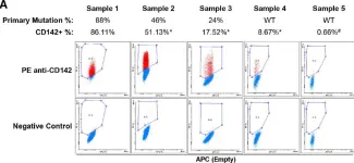
In Cancer Res Commun on 1 April 2023 by Al-Jazrawe, M., Xu, S., et al.
The interaction between neoplastic and stromal cells within a tumor mass plays an important role in cancer biology. However, it is challenging to distinguish between tumor and stromal cells in mesenchymal tumors because lineage-specific cell surface markers typically used in other cancers do not distinguish between the different cell subpopulations. Desmoid tumors consist of mesenchymal fibroblast-like cells driven by mutations stabilizing beta-catenin. Here we aimed to identify surface markers that can distinguish mutant cells from stromal cells to study tumor-stroma interactions. We analyzed colonies derived from single cells from human desmoid tumors using a high-throughput surface antigen screen, to characterize the mutant and nonmutant cells. We found that CD142 is highly expressed by the mutant cell populations and correlates with beta-catenin activity. CD142-based cell sorting isolated the mutant population from heterogeneous samples, including one where no mutation was previously detected by traditional Sanger sequencing. We then studied the secretome of mutant and nonmutant fibroblastic cells. PTX3 is one stroma-derived secreted factor that increases mutant cell proliferation via STAT6 activation. These data demonstrate a sensitive method to quantify and distinguish neoplastic from stromal cells in mesenchymal tumors. It identifies proteins secreted by nonmutant cells that regulate mutant cell proliferation that could be therapeutically.
Distinguishing between neoplastic (tumor) and non-neoplastic (stromal) cells within mesenchymal tumors is particularly challenging, because lineage-specific cell surface markers typically used in other cancers do not differentiate between the different cell subpopulations. Here, we developed a strategy combining clonal expansion with surface proteome profiling to identify markers for quantifying and isolating mutant and nonmutant cell subpopulations in desmoid tumors, and to study their interactions via soluble factors.
© 2023 The Authors; Published by the American Association for Cancer Research.
-
FC/FACS
-
Cancer Research
In Cancer Res Commun on 1 April 2023 by Al-Jazrawe, M., Xu, S., et al.
Fig.4.A

-
FC/FACS
-
Collected and cropped from Cancer Res Commun by CiteAb, provided under a CC-BY license
Image 1 of 1