Compared with long bone that arises from the mesoderm, the major portion of the maxillofacial bones and the front bone of the skull are derived from cranial neural crest cells and undergo intramembranous ossification. Human skeletal stem cells have been identified in embryonic and fetal long bones. Here, we describe a single-cell atlas of the human embryonic mandible and identify a population of cranio-maxillofacial skeletal stem cells (CMSSCs). These CMSSCs are marked by interferon-induced transmembrane protein 5 (IFITM5) and are specifically located around the periosteum of the jawbone and frontal bone. Additionally, these CMSSCs exhibit strong self-renewal and osteogenic differentiation capacities but lower chondrogenic differentiation potency, mediating intramembranous bone formation without cartilage formation. IFITM5+ cells are also observed in the adult jawbone and exhibit functions similar to those of embryonic CMSSCs. Thus, this study identifies CMSSCs that orchestrate the intramembranous ossification of cranio-maxillofacial bones, providing a deeper understanding of cranio-maxillofacial skeletal development and promising seed cells for bone repair.
Product Citations: 76
Identification of human cranio-maxillofacial skeletal stem cells for mandibular development.
In Science Advances on 3 January 2025 by Wang, Z., Wang, K., et al.
-
Stem Cells and Developmental Biology
In Journal of Translational Medicine on 14 August 2024 by Tanzi, A., Buono, L., et al.
Personalized disease models are crucial for evaluating how diseased cells respond to treatments, especially in case of innovative biological therapeutics. Extracellular vesicles (EVs), nanosized vesicles released by cells for intercellular communication, have gained therapeutic interest due to their ability to reprogram target cells. We here utilized urinary podocytes obtained from children affected by steroid-resistant nephrotic syndrome with characterized genetic mutations as a model to test the therapeutic potential of EVs derived from kidney progenitor cells (nKPCs).
EVs were isolated from nKPCs derived from the urine of a preterm neonate. Three lines of urinary podocytes obtained from nephrotic patients' urine and a line of Alport syndrome patient podocytes were characterized and used to assess albumin permeability in response to nKPC-EVs or various drugs. RNA sequencing was conducted to identify commonly modulated pathways after nKPC-EV treatment. siRNA transfection was used to demonstrate the involvement of SUMO1 and SENP2 in the modulation of permeability.
Treatment with the nKPC-EVs significantly reduced permeability across all the steroid-resistant patients-derived and Alport syndrome-derived podocytes. At variance, podocytes appeared unresponsive to standard pharmacological treatments, with the exception of one line, in alignment with the patient's clinical response at 48 months. By RNA sequencing, only two genes were commonly upregulated in nKPC-EV-treated genetically altered podocytes: small ubiquitin-related modifier 1 (SUMO1) and Sentrin-specific protease 2 (SENP2). SUMO1 and SENP2 downregulation increased podocyte permeability confirming the role of the SUMOylation pathway.
nKPCs emerge as a promising non-invasive source of EVs with potential therapeutic effects on podocytes with genetic dysfunction, through modulation of SUMOylation, an important pathway for the stability of podocyte slit diaphragm proteins. Our findings also suggest the feasibility of developing a non-invasive in vitro model for screening regenerative compounds on patient-derived podocytes.
© 2024. The Author(s).
-
FC/FACS
-
Homo sapiens (Human)
In Frontiers in Cell and Developmental Biology on 8 March 2024 by Smolinska, A., Chodkowska, M., et al.
Background: High heterogeneity of mesenchymal stem/stromal cells (MSCs) due to different degrees of differentiation of cell subpopulations poses a considerable challenge in preclinical studies. The cells at a pluripotent-like stage represent a stem cell population of interest for many researchers worldwide, which is worthy of identification, isolation, and functional characterization. In the current study, we asked whether Wharton's jelly-derived MSCs (WJ-MSCs) which express stage-specific embryonic antigen-4 (SSEA-4) can be considered as a pluripotent-like stem cell population. Methods: SSEA-4 expression in different culture conditions was compared and the efficiency of two cell separation methods were assessed: Magnetic Activated Cell Sorting (MACS) and Fluorescence Activated Cell Sorting (FACS). After isolation, SSEA-4+ cells were analyzed for the following parameters: the maintenance of the SSEA-4 antigen expression after cell sorting, stem cell-related gene expression, proliferation potential, clonogenicity, secretome profiling, and the ability to form spheres under 3D culture conditions. Results: FACS allowed for the enrichment of SSEA-4+ cell content in the population that lasted for six passages after sorting. Despite the elevated expression of stemness-related genes, SSEA-4+ cells neither differed in their proliferation and clonogenicity potential from initial and negative populations nor exhibited pluripotent differentiation repertoire. SSEA-4+ cells were observed to form smaller spheroids and exhibited increased survival under 3D conditions. Conclusion: Despite the transient expression of stemness-related genes, our findings could not fully confirm the undifferentiated pluripotent-like nature of the SSEA-4+ WJ-MSC population cultured in vitro.
Copyright © 2024 Smolinska, Chodkowska, Kominek, Janiec, Piwocka, Sulejczak and Sarnowska.
-
Homo sapiens (Human)
Profiling human brain vascular cells using single-cell transcriptomics and organoids.
In Nature Protocols on 1 March 2024 by Crouch, E. E., Diafos, L. N., et al.
Angiogenesis and neurogenesis are functionally interconnected during brain development. However, the study of the vasculature has trailed other brain cell types because they are delicate and of low abundance. Here we describe a protocol extension to purify prenatal human brain endothelial and mural cells with FACS and utilize them in downstream applications, including transcriptomics, culture and organoid transplantation. This approach is simple, efficient and generates high yields from small amounts of tissue. When the experiment is completed within a 24 h postmortem interval, these healthy cells produce high-quality data in single-cell transcriptomics experiments. These vascular cells can be cultured, passaged and expanded for many in vitro assays, including Matrigel vascular tube formation, microfluidic chambers and metabolic measurements. Under these culture conditions, primary vascular cells maintain expression of cell-type markers for at least 3 weeks. Finally, we describe how to use primary vascular cells for transplantation into cortical organoids, which captures key features of neurovascular interactions in prenatal human brain development. In terms of timing, tissue processing and staining requires ~3 h, followed by an additional 3 h of FACS. The transplant procedure of primary, FACS-purified vascular cells into cortical organoids requires an additional 2 h. The time required for different transcriptomic and epigenomic protocols can vary based on the specific application, and we offer strategies to mitigate batch effects and optimize data quality. In sum, this vasculo-centric approach offers an integrated platform to interrogate neurovascular interactions and human brain vascular development.
© 2023. Springer Nature Limited.
Preprint on Research Square on 28 February 2024 by Tanzi, A., Buono, L., et al.
Background: Personalized disease models are crucial for assessing the specific response of diseased cells to drugs, particularly novel biological therapeutics. Extracellular vesicles (EVs), nanosized vesicles released by cells for intercellular communication, have gained therapeutic interest due to their ability to reprogram target cells. We here utilized urinary podocytes obtained from children affected by steroid-resistant nephrotic syndrome with characterized genetic mutations as a model to test the therapeutic potential of EVs derived from kidney progenitor cells. Methods EVs were isolated from kidney progenitor cells (nKPCs) derived from the urine of a preterm neonate. Three lines of urinary podocytes obtained from nephrotic patients' urine and a line of Alport patient podocytes were characterized and used to assess albumin permeability in response to various drugs or to nKPC-EVs. RNA sequencing was conducted to identify commonly modulated pathways. Results Podocytes appeared unresponsive to pharmacological treatments, except for a podocyte line demonstrating responsiveness, in alignment with the patient's clinical response at 48 months. At variance, treatment with the nKPC-EVs was able to significantly reduce permeability in all the steroid-resistant patients-derived podocytes as well as in the line of Alport-derived podocytes. RNA sequencing of nKPC-EV-treated podocytes revealed the common upregulation of two genes (small ubiquitin-related modifier 1 (SUMO1) and Sentrin-specific protease 2 (SENP2)) involved in the SUMOylation pathway, a process recently demonstrated to play a role in slit diaphragm stabilization. Gene ontology analysis on podocyte expression profile highlighted cell-to-cell adhesion as the primary upregulated biological activity in treated podocytes. Conclusions nKPCs emerge as a promising non-invasive source of EVs with potential therapeutic effects on podocyte dysfunction. Furthermore, our findings suggest the possibility of establishing a non-invasive in vitro model for screening regenerative compounds on patient-derived podocytes.
-
Homo sapiens (Human)
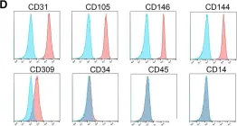
In Stem Cell Res Ther on 22 January 2019 by Gao, K., Kumar, P., et al.
Fig.1.D

-
FC/FACS
-
Homo sapiens (Human)
Collected and cropped from Stem Cell Res Ther by CiteAb, provided under a CC-BY license
Image 1 of 1