Chronic antigenic stimulation can trigger the formation of interleukin 10 (IL-10)-producing T-regulatory type 1 (TR1) cells in vivo. We have recently shown that murine T-follicular helper (TFH) cells are precursors of TR1 cells and that the TFH-to-TR1 cell transdifferentiation process is characterized by the progressive loss and acquisition of opposing transcription factor gene expression programs that evolve through at least one transitional cell stage. Here, we use a broad range of bulk and single-cell transcriptional and epigenetic tools to investigate the epigenetic underpinnings of this process. At the single-cell level, the TFH-to-TR1 cell transition is accompanied by both, downregulation of TFH cell-specific gene expression due to loss of chromatin accessibility, and upregulation of TR1 cell-specific genes linked to chromatin regions that remain accessible throughout the transdifferentiation process, with minimal generation of new open chromatin regions. By interrogating the epigenetic status of accessible TR1 genes on purified TFH and conventional T-cells, we find that most of these genes, including Il10, are already poised for expression at the TFH cell stage. Whereas these genes are closed and hypermethylated in Tconv cells, they are accessible, hypomethylated, and enriched for H3K27ac-marked and hypomethylated active enhancers in TFH cells. These enhancers are enriched for binding sites for the TFH and TR1-associated transcription factors TOX-2, IRF4, and c-MAF. Together, these data suggest that the TR1 gene expression program is genetically imprinted at the TFH cell stage.
© 2024, Garnica et al.
Product Citations: 115
In eLife on 22 November 2024 by Garnica, J., Solé, P., et al.
In IScience on 18 October 2024 by Fahlquist-Hagert, C., Wittenborn, T. R., et al.
Autoantibodies generated in germinal centers (GCs) contribute to the pathogenesis of autoimmune diseases. GCs are controlled by specialized FoxP3+ T-follicular regulatory cells (Tfr), but their role in established autoimmunity is unclear. We generated autoimmune bone marrow chimeras in which Tfr could be specifically ablated by diphtheria toxin. Furthermore, we tracked the clonal persistence and evolution of Tfr populations using Confetti reporters. Ablation of Tfr caused increased early plasma cell output, but longer-term ablation did not increase plasma cell levels beyond those of Tfr-sufficient controls, suggesting that Tfr fail to contain chronic autoreactive GC responses. In agreement, Tfr were robustly induced in early autoreactive GCs but then waned. Moreover, we observed polyclonal Tfr expansion when ablating part of the Tfr subset. Hence, under homeostatic conditions, a polyclonal population of Tfr operates to control autoreactivity by limiting the output of plasma cells from GCs, but in chronic autoimmunity, this mechanism fails.
© 2024 The Author(s).
A TNIP1-driven systemic autoimmune disorder with elevated IgG4.
In Nature Immunology on 1 September 2024 by Medhavy, A., Athanasopoulos, V., et al.
Whole-exome sequencing of two unrelated kindreds with systemic autoimmune disease featuring antinuclear antibodies with IgG4 elevation uncovered an identical ultrarare heterozygous TNIP1Q333P variant segregating with disease. Mice with the orthologous Q346P variant developed antinuclear autoantibodies, salivary gland inflammation, elevated IgG2c, spontaneous germinal centers and expansion of age-associated B cells, plasma cells and follicular and extrafollicular helper T cells. B cell phenotypes were cell-autonomous and rescued by ablation of Toll-like receptor 7 (TLR7) or MyD88. The variant increased interferon-β without altering nuclear factor kappa-light-chain-enhancer of activated B cells signaling, and impaired MyD88 and IRAK1 recruitment to autophagosomes. Additionally, the Q333P variant impaired TNIP1 localization to damaged mitochondria and mitophagosome formation. Damaged mitochondria were abundant in the salivary epithelial cells of Tnip1Q346P mice. These findings suggest that TNIP1-mediated autoimmunity may be a consequence of increased TLR7 signaling due to impaired recruitment of downstream signaling molecules and damaged mitochondria to autophagosomes and may thus respond to TLR7-targeted therapeutics.
© 2024. The Author(s).
-
FC/FACS
-
Mus musculus (House mouse)
-
Immunology and Microbiology
Blockade of OX40/OX40L signaling using anti-OX40L alleviates murine lupus nephritis.
In European Journal of Immunology on 1 August 2024 by Zhao, J., Li, L., et al.
Genetic variants of the OX40 ligand (OX40L) locus are associated with the risk of systemic lupus erythematosus (SLE), it is unclear how the OX40L blockade delays the lupus phenotype. Therefore, we examined the effects of an anti-OX40L antibody in MRL/Lpr mice. Next, we investigated the effect of anti-OX40L on immunosuppression in keyhole limpet hemocyanin-immunized C57BL/6J mice. In vitro treatment of anti-OX40L in CD4+ T and B220+ B cells was used to explore the role of OX40L in the pathogenesis of SLE. Anti-OX40L alleviated murine lupus nephritis, accompanied by decreased production of anti-dsDNA and proteinuria, as well as lower frequencies of splenic T helper (Th) 1 and T-follicular helper cells (Tfh). In keyhole limpet hemocyanin-immunized mice, decreased levels of immunoglobulins and plasmablasts were observed in the anti-OX40L group. Anti-OX40L reduced the number and area of germinal centers. Compared with the control IgG group, anti-OX40L downregulated CD4+ T-cell differentiation into Th1 and Tfh cells and upregulated CD4+ T-cell differentiation into regulatory T cells in vitro. Furthermore, anti-OX40L inhibited toll-like receptor 7-mediated differentiation of antibody-secreting cells and antibody production through the regulation of the SPIB-BLIMP1-XBP1 axis in B cells. These results suggest that OX40L is a promising therapeutic target for SLE.
© 2024 Wiley‐VCH GmbH.
-
Immunology and Microbiology
In Cell Reports Medicine on 16 July 2024 by Perruzza, L., Rezzonico Jost, T., et al.
Environmental enteric dysfunction (EED) is a condition associated with malnutrition that can progress to malabsorption and villous atrophy. Severe EED results in linear growth stunting, slowed neurocognitive development, and unresponsiveness to oral vaccines. Prenatal exposure to malnutrition and breast feeding by malnourished mothers replicates EED. Pups are characterized by deprivation of secretory IgA (SIgA) and altered development of the gut immune system and microbiota. Extracellular ATP (eATP) released by microbiota limits T follicular helper (Tfh) cell activity and SIgA generation in Peyer's patches (PPs). Administration of a live biotherapeutic releasing the ATP-degrading enzyme apyrase to malnourished pups restores SIgA levels and ameliorates stunted growth. SIgA is instrumental in improving the growth and intestinal immune competence of mice while they are continuously fed a malnourished diet. The analysis of microbiota composition suggests that amplification of endogenous SIgA may exert a dominant function in correcting malnourishment dysbiosis and its consequences on host organisms, irrespective of the actual microbial ecology.
Copyright © 2024 The Author(s). Published by Elsevier Inc. All rights reserved.
-
Mus musculus (House mouse)
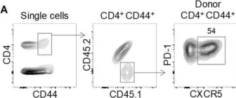
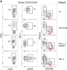
In Front Immunol on 29 June 2018 by Danelli, L., Donnarumma, T., et al.
Fig.1.A

-
FC/FACS
-
Collected and cropped from Front Immunol by CiteAb, provided under a CC-BY license
Image 1 of 7
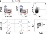
In Front Immunol on 29 June 2018 by Danelli, L., Donnarumma, T., et al.
Fig.1.B

-
FC/FACS
-
Collected and cropped from Front Immunol by CiteAb, provided under a CC-BY license
Image 1 of 7
In Front Immunol on 29 June 2018 by Danelli, L., Donnarumma, T., et al.
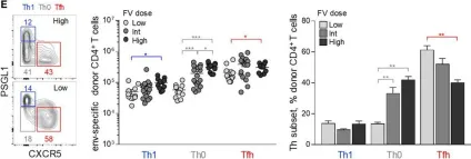
Fig.1.E

-
FC/FACS
-
Collected and cropped from Front Immunol by CiteAb, provided under a CC-BY license
Image 1 of 7
In Front Immunol on 29 June 2018 by Danelli, L., Donnarumma, T., et al.
Fig.3.G

-
FC/FACS
-
Collected and cropped from Front Immunol by CiteAb, provided under a CC-BY license
Image 1 of 7
In Front Immunol on 29 June 2018 by Danelli, L., Donnarumma, T., et al.
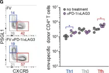
Fig.5.A

-
FC/FACS
-
Collected and cropped from Front Immunol by CiteAb, provided under a CC-BY license
Image 1 of 7
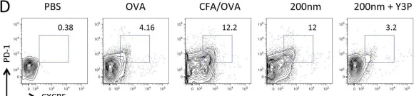
In Elife on 10 August 2015 by Benson, R. A., MacLeod, M. K., et al.
Fig.7.D

-
FC/FACS
-
Collected and cropped from Elife by CiteAb, provided under a CC-BY license
Image 1 of 7
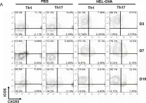
In PLoS One on 21 November 2012 by Patakas, A., Benson, R. A., et al.
Fig.6.A

-
FC/FACS
-
Mus musculus (House mouse)
Collected and cropped from PLoS One by CiteAb, provided under a CC-BY license
Image 1 of 7