Galectin-3 is an endogenous lectin which binds mainly to β-galactosides on the cell surface and extracellular matrix (ECM) glycoproteins. In the thymus, this lectin is constitutively expressed, being involved in thymocyte adhesion, migration, and death. Galectin-3 has been related to type 1 diabetes, an autoimmune disease characterized by pancreatic β-cell destruction mediated by autoreactive T lymphocytes. Non-obese diabetic (NOD) mice represent a suitable model to study type 1 diabetes, as they develop the disease like humans. We previously described important thymic alterations in these animals such as the development of giant perivascular spaces (PVS), characterized by the retention of T and B cells, intermingled with an ECM network, and associated with a defect in the expression of the fibronectin receptor VLA-5 and reduced sphingosine-1-phosphate receptor expression on developing thymocytes. In order to investigate galectin-3 expression in thymic microenvironmental cells and verify its interaction with cells and ECM molecules in PVS, we performed immunofluorescence following colocalization analysis in the thymic parenchyma of pre-diabetic NOD mice by confocal microscopy. In addition, thymocyte migration assays were performed to evaluate the effect of galectin-3 on NOD thymocyte migration. Herein, we showed a significant enhancement of colocalization with cortical and medullary thymic epithelial cells in NOD mice, as compared to controls. In the giant PVS of these animals, we observed a heterogeneous distribution of galectin-3, predominantly found in clusters of B lymphocytes and dendritic cells. Functionally, NOD thymocyte migratory response towards galectin-3 was impaired and a similar decrease was seen in transendothelial thymocyte migration. Taken together, our data provide the histological and functional background for a potential defective thymocyte migration involving galectin-3, thus placing this molecule as a further player in the intrathymic disturbances observed in pre-diabetic NOD mice.
Copyright © 2024 Ramos, Ventura, Lemos, Chammas, Savino, Carvalho-Pinto, Mendes-da-Cruz and Villa-Verde.
Product Citations: 675
In Frontiers in Endocrinology on 4 November 2024 by Ramos, T. D. P., Ventura, A. L. M., et al.
-
Mus musculus (House mouse)
-
Endocrinology and Physiology
Oral reovirus reshapes the gut microbiome and enhances antitumor immunity in colon cancer.
In Nature Communications on 22 October 2024 by Lee, W. S., Lee, S. J., et al.
The route of oncolytic virotherapy is pivotal for immunotherapeutic efficacy in advanced cancers. In this preclinical study, an oncolytic reovirus (RC402) is orally administered to induce antitumor immunity. Oral reovirus treatment shows no gross toxicities and effectively suppresses multifocal tumor lesions. Orally administered reovirus interacts with the host immune system in the Peyer's patch of the terminal ileum, increases IgA+ antibody-secreting cells in the lamina propria through MAdCAM-1+ blood vessels, and reshapes the gut microbiome. Oral reovirus promotes antigen presentation, type I/II interferons, and T cell activation within distant tumors, but does not reach or directly infect tumor cells beyond the gastrointestinal tract. In contrast to intratumoral reovirus injection, the presence of the gut microbiome, Batf3+ dendritic cells, type I interferons, and CD8+ T cells are indispensable for orally administered reovirus-induced antitumor immunity. Oral reovirus treatment is most effective when combined with αPD-1(L1) and/or αCTLA-4, leading to complete colon tumor regression and protective immune memory. Collectively, oral reovirus virotherapy is a feasible and effective immunotherapeutic strategy in preclinical studies.
© 2024. The Author(s).
-
IHC-Frozen
-
IHC
-
Mus musculus (House mouse)
-
Cancer Research
-
Immunology and Microbiology
In Communications Biology on 5 February 2024 by Petrova, E., López-Gay, J. M., et al.
Netherton syndrome (NS) is a rare skin disease caused by loss-of-function mutations in the serine peptidase inhibitor Kazal type 5 (SPINK5) gene. Disease severity and the lack of efficacious treatments call for a better understanding of NS mechanisms. Here we describe a novel and viable, Spink5 conditional knock-out (cKO) mouse model, allowing to study NS progression. By combining transcriptomics and proteomics, we determine a disease molecular profile common to mouse models and NS patients. Spink5 cKO mice and NS patients share skin barrier and inflammation signatures defined by up-regulation and increased activity of proteases, IL-17, IL-36, and IL-20 family cytokine signaling. Systemic inflammation in Spink5 cKO mice correlates with disease severity and is associated with thymic atrophy and enlargement of lymph nodes and spleen. This systemic inflammation phenotype is marked by neutrophils and IL-17/IL-22 signaling, does not involve primary T cell immunodeficiency and is independent of bacterial infection. By comparing skin transcriptomes and proteomes, we uncover several putative substrates of tissue kallikrein-related proteases (KLKs), demonstrating that KLKs can proteolytically regulate IL-36 pro-inflammatory cytokines. Our study thus provides a conserved molecular framework for NS and reveals a KLK/IL-36 signaling axis, adding new insights into the disease mechanisms and therapeutic targets.
© 2024. The Author(s).
-
Mus musculus (House mouse)
Lineage-specific 3D genome organization is assembled at multiple scales by IKAROS.
In Cell on 22 November 2023 by Hu, Y., Salgado Figueroa, D., et al.
A generic level of chromatin organization generated by the interplay between cohesin and CTCF suffices to limit promiscuous interactions between regulatory elements, but a lineage-specific chromatin assembly that supersedes these constraints is required to configure the genome to guide gene expression changes that drive faithful lineage progression. Loss-of-function approaches in B cell precursors show that IKAROS assembles interactions across megabase distances in preparation for lymphoid development. Interactions emanating from IKAROS-bound enhancers override CTCF-imposed boundaries to assemble lineage-specific regulatory units built on a backbone of smaller invariant topological domains. Gain of function in epithelial cells confirms IKAROS' ability to reconfigure chromatin architecture at multiple scales. Although the compaction of the Igκ locus required for genome editing represents a function of IKAROS unique to lymphocytes, the more general function to preconfigure the genome to support lineage-specific gene expression and suppress activation of extra-lineage genes provides a paradigm for lineage restriction.
Published by Elsevier Inc.
-
Mus musculus (House mouse)
In eLife on 9 August 2023 by Wang, Y., Wu, T., et al.
Tumor progression locus 2 (TPL2) (MAP3K8) is a central signaling node in the inflammatory response of peripheral immune cells. We find that TPL2 kinase activity modulates microglial cytokine release and is required for microglia-mediated neuron death in vitro. In acute in vivo neuroinflammation settings, TPL2 kinase activity regulates microglia activation states and brain cytokine levels. In a tauopathy model of chronic neurodegeneration, loss of TPL2 kinase activity reduces neuroinflammation and rescues synapse loss, brain volume loss, and behavioral deficits. Single-cell RNA sequencing analysis indicates that protection in the tauopathy model was associated with reductions in activated microglia subpopulations as well as infiltrating peripheral immune cells. Overall, using various models, we find that TPL2 kinase activity can promote multiple harmful consequences of microglial activation in the brain including cytokine release, iNOS (inducible nitric oxide synthase) induction, astrocyte activation, and immune cell infiltration. Consequently, inhibiting TPL2 kinase activity could represent a potential therapeutic strategy in neurodegenerative conditions.
© 2023, Wang et al.
-
Mus musculus (House mouse)
-
Immunology and Microbiology
-
Neuroscience
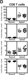
In PLoS One on 6 February 2017 by Tan, S. Y., Chowdhury, S., et al.
Fig.1.D

-
FC/FACS
-
Collected and cropped from PLoS One by CiteAb, provided under a CC-BY license
Image 1 of 4
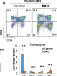
In Oncotarget on 9 August 2016 by Goetz, B., An, W., et al.
Fig.3.B

-
FC/FACS
-
Collected and cropped from Oncotarget by CiteAb, provided under a CC-BY license
Image 1 of 4
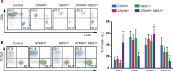
In PLoS Genet on 1 November 2015 by Prochazkova, J., Sakaguchi, S., et al.
Fig.5.D

-
FC/FACS
-
Collected and cropped from PLoS Genet by CiteAb, provided under a CC-BY license
Image 1 of 4
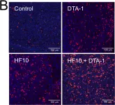
In PLoS One on 12 August 2014 by Ishihara, M., Seo, N., et al.
Fig.2.B

-
ICC
-
Mus musculus (House mouse)
Collected and cropped from PLoS One by CiteAb, provided under a CC-BY license
Image 1 of 4