Highly conserved homeobox genes are closely related to bone formation during embryogenesis, while their role in adult bone resorption remains unclear. In this study, we found that the homeobox gene MSX2 actively participates bone metabolism. Myeloid-specific Msx2 deficiency safeguards bone mass under physiological and pathological conditions. Loss of Msx2 acts as a "brake" in the fusion fate of osteoclasts, resulting in a larger population of pre-osteoclasts. Pre-osteoclasts secrete platelet-derived growth factor-BB (PDGF-BB), which promotes angiogenesis-mediated bone formation. Mechanistically, MSX2 directly binds to the vital osteoclastogenic transcription factor PU.1 and protects it from FBXW7-mediated ubiquitination degradation. Msx2 and Fbxw7 double knockout mitigated the protective effect of MSX2 deficiency on bone mass. Finally, we identified a natural compound, morusinol, that specifically destroys the combination of MSX2 and PU.1, promoting PU.1 degradation and attenuating ovariectomy-induced bone loss. Overall, our results demonstrate that targeting Msx2 is a promising anabolic therapy for osteoporosis.
© 2025. The Author(s).
Product Citations: 131
In Nature Communications on 6 August 2025 by Ma, Q., Wang, S., et al.
In IScience on 18 July 2025 by Lu, L., Xu, K., et al.
The pathogenic factors of inflammatory bowel disease (IBD) are very complex, and further investigation of its pathogenesis is necessary. Some polar proteins have been reported to perform functions other than maintaining cell structure. In this study, we reported that the polar protein AF6 expressed by intestinal epithelial cells (IEC) can regulate the expression of MHCII in epithelial cells, thus affecting the cross dialogue between IECs and immune cells, and promoting the secretion of IgA by B cells to play a protective function. IgA supplementation can also improve the severe colitis phenotype caused by AF6 loss. Our study provides an understanding of the regulatory mechanisms of the intestinal epithelial expression of MHC II and its important role in colitis, which will aid in treatment and intervention.
© 2025 The Authors.
-
Immunology and Microbiology
Obesity drives depot-specific vascular remodeling in male white adipose tissue.
In Nature Communications on 25 June 2025 by Hasan, S. S., John, D., et al.
Obesity-driven pathological expansion of white adipose tissue (WAT) is a key driver of endothelial dysfunction. However, early vascular alterations associated with over-nutrition also serve to exacerbate WAT dysfunction. Here, we conduct a single-cell transcriptomic analysis of WAT endothelium to delineate endothelial heterogeneity and elucidate vascular alterations and its consequence in a male murine model of obesity. We demarcate depot-specific differences in subcutaneous (sWAT) and visceral WAT (vWAT) endothelium through in sillico analysis and further corroboration of our findings. Moreover, we identify a sWAT-specific fenestrated endothelial cell (EC) subtype, which declines in obese conditions. Utilizing systemic anti-VEGFA blockade and genetic Vegfa manipulation, we demonstrate that VEGFA is necessary for maintaining fenestration in sWAT. Additionally, we detect this fenestrated EC subtype in male human WAT, which undergoes reduction in individuals with obesity. Collectively, this atlas serves as a valuable tool for future studies to decipher the functional significance of different WAT EC subtypes.
© 2025. The Author(s).
Cbl-b inhibitor NX-1607 activates MAPK/ERK signaling pathway and enhances T-cell activation.
In Journal for Immunotherapy of Cancer on 30 May 2025 by Zhu, W., Lu, S., et al.
Backgroud: The E3 ubiquitin ligase casitas B lymphoma-b (Cbl-b) is pivotal in modulating immune responses by attenuating T-cell activation and cytokine production. Inhibiting Cbl-b presents a potential therapeutic strategy in immuno-oncology by enhancing immune activity. Methods: A rapid Homogeneous Time-Resolved Fluorescence (HTRF) assay was employed to evaluate the inhibitory efficacy of NX-1607 on Cbl-b. The effects of NX-1607 on T cell activation, cytokine production, and proliferation were characterized invitro using primary T cells and Jurkat T cells. A drug combination screening was performed utilizing the CD69 marker via flow cytometry to dentify signaling pathways involved in T cell activation by NX-1607. CRISPR/Cas9 technology was used to knock out PLCG1 and MAPK3/1 in Jurkat T cells, followed by the detection of p-PLCγ1 and p-ERK1/2 though Western blotting. The antitumor efficacy of NX-1607 was assessed in a murine model of A20 B-cell lymphoma using BALB/c mice, with subsequent flow cytometry analysis conducted to examine the phenotype of tumor-infiltrating lymphocytes (TILs). Results: Our data show that NX-1607 effectively inhibits Cbl-b activity at low nanomolar levels, boosting PLCγ1 and HCSL1 phosphorylation, activating MAPK/ERK signaling, and elevating CD69 expression. Inhibiting PLCγ1 and ERK1/2 significantly reduces NX-1607's effect on T-cell activation. Oral administration of NX-1607 notably decreases tumor growth in the A20 B-cell lymphoma model, with immunophenotyping analyses of tumor-infiltrating lymphocytes revealing increased CD3+, CD4+, and CD8+ T cells in treated tumors. Furthermore, our results demonstrate that treatment with NX-1607 results in increased levels of phosphorylated PLCγ1 and ERK1/2 in circulating T cells. Conclusion: Taken together, these findings imply that the inhibition of Cbl-b by NX-1607 may enhance the activation of the MAPK/ERK signaling pathway, thereby sustaining T-cell activation. This provides compelling evidence for the molecular mechanism of NX-1607, underscoring the pivotal role of Cbl-b in controlling signal strength in T-cell activation after T-cell receptor (TCR) engagement.
© Author(s) (or their employer(s)) 2025. Re-use permitted under CC BY-NC. No commercial re-use. See rights and permissions. Published by BMJ Group.
-
FC/FACS
-
Immunology and Microbiology
In CNS Neuroscience Therapeutics on 1 May 2025 by Dai, X., Zhang, X., et al.
Following ischemic stroke, peripheral immune cell infiltration is characterized by myeloid cell predominance in the acute phase and lymphoid cell infiltration in the subacute to chronic phases. Endothelial cells, as a critical interface between the peripheral circulation and the brain, upregulate adhesion molecules to facilitate immune cell infiltration. However, it remains unclear whether endothelial cells exhibit functional differences at different stages after ischemic stroke and how these differences affect immune cell infiltration.
We performed single-cell RNA sequencing on peripheral immune and endothelial cells from Sham and middle cerebral artery occlusion (MCAO) mice at 3 and 14 days post-MCAO. Subsequent analysis of the sequencing data, combined with flow cytometry and immunofluorescence staining, was used to investigate the relationship between endothelial cell changes at different stages of stroke and immune cell infiltration.
We observed that the infiltration capacity of peripheral immune cells did not significantly increase at different stages after MCAO. However, endothelial cells underwent significant changes. By Day 3 post-MCAO, there was an increased proportion of venous endothelial cells with enhanced angiogenesis and adhesion functions. In this acute phase, newly formed venous endothelial cells with high expression of the adhesion molecule ICAM-1 were observed, promoting the infiltration of myeloid cells and NKT cells. From the acute to chronic phases, endothelial angiogenesis gradually decreased, accompanied by a marked increase in antigen presentation function. At 14 days post-MCAO, an increased proportion of VCAM-1-expressing venous endothelial cells was observed, potentially facilitating the infiltration of T cells and a subset of neutrophils. Furthermore, we discovered that the differential changes in venous endothelial cells at different stages after MCAO may be driven by distinct differentiation and proliferation patterns regulated by different signaling pathways.
Our study highlights that the differential expression of adhesion molecules and functional changes in endothelial cells at distinct stages after ischemic stroke may regulate the infiltration patterns of peripheral immune cells.
© 2025 The Author(s). CNS Neuroscience & Therapeutics published by John Wiley & Sons Ltd.
-
Cardiovascular biology
-
Immunology and Microbiology
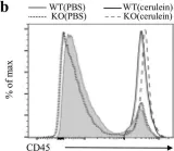
In Sci Rep on 7 November 2018 by Quan, S., Principe, D. R., et al.
Fig.2.B

-
FC/FACS
-
Mus musculus (House mouse)
Collected and cropped from Sci Rep by CiteAb, provided under a CC-BY license
Image 1 of 1