CD4+ T cell activation is driven by five-module receptor complexes. The T cell receptor (TCR) is the receptor module that binds composite surfaces of peptide antigens embedded within MHCII molecules (pMHCII). It associates with three signaling modules (CD3γε, CD3δε, and CD3ζζ) to form TCR-CD3 complexes. CD4 is the coreceptor module. It reciprocally associates with TCR-CD3-pMHCII assemblies on the outside of a CD4+ T cells and with the Src kinase, LCK, on the inside. Previously, we reported that the CD4 transmembrane GGXXG and cytoplasmic juxtamembrane (C/F)CV+C motifs found in eutherian (placental mammal) CD4 have constituent residues that evolved under purifying selection (Lee et al., 2022). Expressing mutants of these motifs together in T cell hybridomas increased CD4-LCK association but reduced CD3ζ, ZAP70, and PLCγ1 phosphorylation levels, as well as IL-2 production, in response to agonist pMHCII. Because these mutants preferentially localized CD4-LCK pairs to non-raft membrane fractions, one explanation for our results was that they impaired proximal signaling by sequestering LCK away from TCR-CD3. An alternative hypothesis is that the mutations directly impacted signaling because the motifs normally play an LCK-independent role in signaling. The goal of this study was to discriminate between these possibilities. Using T cell hybridomas, our results indicate that: intracellular CD4-LCK interactions are not necessary for pMHCII-specific signal initiation; the GGXXG and (C/F)CV+C motifs are key determinants of CD4-mediated pMHCII-specific signal amplification; the GGXXG and (C/F)CV+C motifs exert their functions independently of direct CD4-LCK association. These data provide a mechanistic explanation for why residues within these motifs are under purifying selection in jawed vertebrates. The results are also important to consider for biomimetic engineering of synthetic receptors.
© 2023, Lee et al.
Product Citations: 18
In eLife on 19 April 2024 by Lee, M. S., Tuohy, P. J., et al.
-
Immunology and Microbiology
Construction of a T cell receptor signaling range for spontaneous development of autoimmune disease.
In The Journal of Experimental Medicine on 6 February 2023 by Tanaka, A., Maeda, S., et al.
Thymic selection and peripheral activation of conventional T (Tconv) and regulatory T (Treg) cells depend on TCR signaling, whose anomalies are causative of autoimmunity. Here, we expressed in normal mice mutated ZAP-70 molecules with different affinities for the CD3 chains, or wild type ZAP-70 at graded expression levels under tetracycline-inducible control. Both manipulations reduced TCR signaling intensity to various extents and thereby rendered those normally deleted self-reactive thymocytes to become positively selected and form a highly autoimmune TCR repertoire. The signal reduction more profoundly affected Treg development and function because their TCR signaling was further attenuated by Foxp3 that physiologically repressed the expression of TCR-proximal signaling molecules, including ZAP-70, upon TCR stimulation. Consequently, the TCR signaling intensity reduced to a critical range generated pathogenic autoimmune Tconv cells and concurrently impaired Treg development/function, leading to spontaneous occurrence of autoimmune/inflammatory diseases, such as autoimmune arthritis and inflammatory bowel disease. These results provide a general model of how altered TCR signaling evokes autoimmune disease.
© 2022 Tanaka et al.
-
FC/FACS
-
Mus musculus (House mouse)
-
Immunology and Microbiology
Naïve arthritogenic SKG T cells have a defect in anergy and a repertoire pruned by superantigen
Preprint on BioRxiv : the Preprint Server for Biology on 16 January 2022 by Ashouri, J. F., McCarthy, E., et al.
How autoreactive CD4 T cells develop to cause rheumatoid arthritis remains unknown. We used a reporter for antigen-receptor signaling in the SKG autoimmune arthritis model to profile a T cell subpopulation enriched for arthritogenic naïve CD4 T cells before arthritis onset by bulk and single cell RNA and T cell antigen-receptor (TCR) sequencing. Our analyses reveal that despite their impaired proximal TCR signaling, a subset of SKG naïve CD4 T cells that have recently encountered endogenous antigen upregulate gene programs associated with positive regulation of T cell activation and cytokine signaling at higher levels than wild type cells in the pre-disease state. These arthritogenic cells also induce genes associated with negative regulation of T cell activation but do so less efficiently than wild type cells. Furthermore, their TCR sequences exhibit a previously unrecognized biased peripheral TCR Vβ repertoire likely driven by endogenous viral superantigens. These particular Vβs, known to recognize endogenous mouse mammary tumor virus (MMTV) superantigen, are further expanded in arthritic joints. Our results demonstrate that autoreactive naïve CD4 T cells which recognize endogenous viral superantigens are poised to cause disease by their altered transcriptome. h4>Summary blurb/h4> Self-reactive SKG T cells that escaped negative selection harbor an independent defect in anergy that, together with chronic antigen stimulation, sets the stage for disease. Moreover, we propose a novel role for endogenous mouse mammary tumor virus (MMTV) superantigen in promoting arthritogenic T cell responses.
-
Immunology and Microbiology
In Frontiers in Immunology on 27 April 2021 by Prasad, M., Wojciech, L., et al.
Deletion of the gene for Themis affects T cell selection in the thymus, which would be expected to affect the TCR repertoire. We found an increased proportion of cells expressing Vα3.2 (TRAV9N-3) in the peripheral CD8+ T cell population in mice with germline Themis deficiency. Analysis of the TCRα repertoire indicated it was generally reduced in diversity in the absence of Themis, whereas the diversity of sequences using the TRAV9N-3 V-region element was increased. In wild type mice, Vα3.2+ cells showed higher CD5, CD6 and CD44 expression than non-Vα3-expressing cells, and this was more marked in cells from Themis-deficient mice. This suggested a virtual memory phenotype, as well as a stronger response to self-pMHC. The Vα3.2+ cells responded more strongly to IL-15, as well as showing bystander effector capability in a Listeria infection. Thus, the unusually large population of Vα3.2+ CD8+ T cells found in the periphery of Themis-deficient mice reflects not only altered thymic selection, but also allowed identification of a subset of bystander-competent cells that are also present in wild-type mice.
Copyright © 2021 Prasad, Wojciech, Brzostek, Hu, Chua, Tung, Yap, Rybakin and Gascoigne.
-
Mus musculus (House mouse)
-
Immunology and Microbiology
In Immunity, Inflammation and Disease on 1 June 2018 by Omodho, B., Miao, T., et al.
Impaired proliferation and production of IL2 are the hallmarks of experimental T cell tolerance. However, in most autoimmune diseases, auto-reactive T cells do not display hyper proliferation, but inflammatory phenotypes.
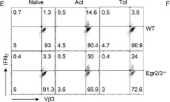
We have now demonstrated that the transcription factors Egr2 and 3 are important for the control of inflammatory cytokine production by tolerant T cells, but not for tolerance induction.
In the absence of Egr2 and 3, T cell tolerance, as measured by impaired proliferation and production of IL2, can still be induced, but tolerant T cells produced high levels of inflammatory cytokines. Egr2 and 3 regulate expression of differentiation repressors and directly inhibit T-bet function in T cells. Indeed, decreased expression of differentiation repressors, such as Id3 and Tcf1, and increased expression of inflammatory transcription factors, such as RORγt and Bhlhe40 were found in Egr2/3 deficient T cells under tolerogenic conditions. In addition, T-bet was co-expressed with Egr2 in tolerant T cells and Egr2/3 defects leads to production of high levels of IFNγ in tolerant T cells.
Our findings demonstrated that despite impaired proliferation and IL2 production, tolerant T cells can display inflammatory responses in response to antigen stimulation and this is controlled at least partly by Egr2 and 3.
© 2018 The Authors. Immunity, Inflammation and Disease Published by John Wiley & Sons Ltd.
-
FC/FACS
-
Mus musculus (House mouse)
-
Biochemistry and Molecular biology
-
Immunology and Microbiology
In Immun Inflamm Dis on 1 June 2018 by Omodho, B., Miao, T., et al.
Fig.5.E

-
FC/FACS
-
Mus musculus (House mouse)
Collected and cropped from Immun Inflamm Dis by CiteAb, provided under a CC-BY license
Image 1 of 1