Atherosclerosis, characterized by the accumulation of lipid plaques on the inner walls of arteries, is the leading cause of heart attack, stroke and severe ischemic injuries. Senescent cells have been found to accumulate within atherosclerotic lesions and contribute to the progression of atherosclerosis. In our previous study, we discovered that suppressing Larp7 accelerates senescence by inhibiting Sirt1 activity, resulting in increased atherosclerosis in high-fat diet (HFD) fed and ApoE deficient (ApoEKO) mice. However, there has been no direct evidence demonstrating Larp7 per se could attenuate atherosclerosis. To this end, we generated a tetO-controlled and Cre-activated Larp7 gain-of-function mouse. Through RT-PCR and western blotting, we confirmed Larp7 overexpression in the aortas of HFD-fed ApoEKO; Larp7tetO mice. Larp7 overexpression led to increased Sirt1 activity and decreased cellular senescence signals mediated by p53/p65 in the aortas. Additionally, Larp7 overexpression reduced the presence of p16-positive senescent cells in the aortic lesions. Furthermore, Larp7 overexpression resulted in a decrease in pro-inflammatory macrophages and SASP factors. Consequently, Larp7 overexpression led to a reduction in the area of atherosclerotic lesions in HFD-fed ApoEKO; Larp7tetO mice. In summary, our study provides evidence that Larp7 overexpression holds promise as an approach to inhibit cellular senescence and prevent atherosclerosis.
© 2024 The Author(s). Journal of Cellular and Molecular Medicine published by Foundation for Cellular and Molecular Medicine and John Wiley & Sons Ltd.
Product Citations: 117
LARP7 overexpression alleviates aortic senescence and atherosclerosis.
In Journal of Cellular and Molecular Medicine on 1 June 2024 by Yang, P., Wu, S., et al.
-
Mus musculus (House mouse)
-
Biochemistry and Molecular biology
Ligand-dependent interactions between SR-B1 and S1PR1 in macrophages and atherosclerotic plaques.
In Journal of Lipid Research on 1 May 2024 by Bassila, C., Kluck, G. E. G., et al.
HDLs carry sphingosine-1-phosphate (S1P) and stimulate signaling pathways in different cells including macrophages and endothelial cells, involved in atherosclerotic plaque development. HDL signaling via S1P relies on the HDL receptor scavenger receptor class B, type I (SR-B1) and the sphingosine-1-phosphate receptor 1 (S1PR1), which interact when both are heterologously overexpressed in the HEK293 cell line. In this study, we set out to test if SR-B1 and S1PR1 interacted in primary murine macrophages in culture and atherosclerotic plaques. We used knock-in mice that endogenously expressed S1PR1 tagged with eGFP-(S1pr1eGFP/eGFP mice), combined with proximity ligation analysis to demonstrate that HDL stimulates the physical interaction between SR-B1 and S1PR1 in primary macrophages, that this is dependent on HDL-associated S1P and can be blocked by an inhibitor of SR-B1's lipid transfer activity or an antagonist of S1PR1. We also demonstrate that a synthetic S1PR1-selective agonist, SEW2871, stimulates the interaction between SR-B1 and S1PR1 and that this was also blocked by an inhibitor of SR-B1's lipid transport activity. Furthermore, we detected abundant SR-B1/S1PR1 complexes in atherosclerotic plaques of S1pr1eGFP/eGFP mice that also lacked apolipoprotein E. Treatment of mice with the S1PR1 antagonist, Ex26, for 12 h disrupted the SR-B1-S1PR1 interaction in atherosclerotic plaques. These findings demonstrate that SR-B1 and S1PR1 form ligand-dependent complexes both in cultured primary macrophages and within atherosclerotic plaques in mice and provide mechanistic insight into how SR-B1 and S1PR1 participate in mediating HDL signaling to activate atheroprotective responses in macrophages.
Copyright © 2024 The Authors. Published by Elsevier Inc. All rights reserved.
-
Mus musculus (House mouse)
Selenoprotein deficiency disorder predisposes to aortic aneurysm formation.
In Nature Communications on 2 December 2023 by Schoenmakers, E., Marelli, F., et al.
Aortic aneurysms, which may dissect or rupture acutely and be lethal, can be a part of multisystem disorders that have a heritable basis. We report four patients with deficiency of selenocysteine-containing proteins due to selenocysteine Insertion Sequence Binding Protein 2 (SECISBP2) mutations who show early-onset, progressive, aneurysmal dilatation of the ascending aorta due to cystic medial necrosis. Zebrafish and male mice with global or vascular smooth muscle cell (VSMC)-targeted disruption of Secisbp2 respectively show similar aortopathy. Aortas from patients and animal models exhibit raised cellular reactive oxygen species, oxidative DNA damage and VSMC apoptosis. Antioxidant exposure or chelation of iron prevents oxidative damage in patient's cells and aortopathy in the zebrafish model. Our observations suggest a key role for oxidative stress and cell death, including via ferroptosis, in mediating aortic degeneration.
© 2023. The Author(s).
-
IHC
-
Mus musculus (House mouse)
In European Heart Journal on 1 August 2023 by Meng, Z., Zhang, S., et al.
Blood eosinophil count and eosinophil cationic protein (ECP) concentration are risk factors of cardiovascular diseases. This study tested whether and how eosinophils and ECP contribute to vascular calcification and atherogenesis.
Immunostaining revealed eosinophil accumulation in human and mouse atherosclerotic lesions. Eosinophil deficiency in ΔdblGATA mice slowed atherogenesis with increased lesion smooth muscle cell (SMC) content and reduced calcification. This protection in ΔdblGATA mice was muted when mice received donor eosinophils from wild-type (WT), Il4-/-, and Il13-/- mice or mouse eosinophil-associated-ribonuclease-1 (mEar1), a murine homologue of ECP. Eosinophils or mEar1 but not interleukin (IL) 4 or IL13 increased the calcification of SMC from WT mice but not those from Runt-related transcription factor-2 (Runx2) knockout mice. Immunoblot analyses showed that eosinophils and mEar1 activated Smad-1/5/8 but did not affect Smad-2/3 activation or expression of bone morphogenetic protein receptors (BMPR-1A/1B/2) or transforming growth factor (TGF)-β receptors (TGFBR1/2) in SMC from WT and Runx2 knockout mice. Immunoprecipitation showed that mEar1 formed immune complexes with BMPR-1A/1B but not TGFBR1/2. Immunofluorescence double-staining, ligand binding, and Scatchard plot analysis demonstrated that mEar1 bound to BMPR-1A and BMPR-1B with similar affinity. Likewise, human ECP and eosinophil-derived neurotoxin (EDN) also bound to BMPR-1A/1B on human vascular SMC and promoted SMC osteogenic differentiation. In a cohort of 5864 men from the Danish Cardiovascular Screening trial and its subpopulation of 394 participants, blood eosinophil counts and ECP levels correlated with the calcification scores of different arterial segments from coronary arteries to iliac arteries.
Eosinophils release cationic proteins that can promote SMC calcification and atherogenesis using the BMPR-1A/1B-Smad-1/5/8-Runx2 signalling pathway.
© The Author(s) 2023. Published by Oxford University Press on behalf of the European Society of Cardiology.
-
Mus musculus (House mouse)
-
Cardiovascular biology
cGAS-STING drives ageing-related inflammation and neurodegeneration.
In Nature on 1 August 2023 by Gulen, M. F., Samson, N., et al.
Low-grade inflammation is a hallmark of old age and a central driver of ageing-associated impairment and disease1. Multiple factors can contribute to ageing-associated inflammation2; however, the molecular pathways that transduce aberrant inflammatory signalling and their impact in natural ageing remain unclear. Here we show that the cGAS-STING signalling pathway, which mediates immune sensing of DNA3, is a critical driver of chronic inflammation and functional decline during ageing. Blockade of STING suppresses the inflammatory phenotypes of senescent human cells and tissues, attenuates ageing-related inflammation in multiple peripheral organs and the brain in mice, and leads to an improvement in tissue function. Focusing on the ageing brain, we reveal that activation of STING triggers reactive microglial transcriptional states, neurodegeneration and cognitive decline. Cytosolic DNA released from perturbed mitochondria elicits cGAS activity in old microglia, defining a mechanism by which cGAS-STING signalling is engaged in the ageing brain. Single-nucleus RNA-sequencing analysis of microglia and hippocampi of a cGAS gain-of-function mouse model demonstrates that engagement of cGAS in microglia is sufficient to direct ageing-associated transcriptional microglial states leading to bystander cell inflammation, neurotoxicity and impaired memory capacity. Our findings establish the cGAS-STING pathway as a driver of ageing-related inflammation in peripheral organs and the brain, and reveal blockade of cGAS-STING signalling as a potential strategy to halt neurodegenerative processes during old age.
© 2023. The Author(s).
-
Immunology and Microbiology
-
Neuroscience
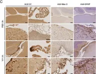
In Acta Neuropathol Commun on 7 March 2014 by Rothhammer, V., Muschaweckh, A., et al.
Fig.1.C

-
IHC
-
Collected and cropped from Acta Neuropathol Commun by CiteAb, provided under a CC-BY license
Image 1 of 1