Abstract Lipedema is a hereditary disorder characterized by excessive accumulation of subcutaneous adipose tissue in the limbs. The genetic causes and mechanisms underlying abnormal adipocyte expansion in lipedema, however, remain unknown. Here, we identify compound heterozygous mutations in the PROC gene in three lipedema patients from two unrelated consanguineous families. In vitro studies demonstrate the wild-type Protein C (PC), encoded by PROC, plays an inhibitory role in adipogenesis; conversely, the identified PC mutants, p.R271Q and p.R272H, fail to inhibit this process. In mice, the receptor of PC (PROCR) marks adipocyte progenitors, and conditional deletion of PROCR in these cells leads to an increased number of newborn adipocytes within white adipose tissue (WAT). Transcriptomic analysis alongside chemical blockage tests identifies HIF-1α as a primary downstream transcription factor mediating PC–PROCR signaling in adipogenesis. Furthermore, adipose biopsy samples from the patients’ thighs exhibit hyperplastic expansion of adipocytes, while single-nucleus RNA sequencing confirms increased adipogenic capacity and down-regulated HIF-1α activity in affected subjects. These findings establish PROC as the first causal gene for human lipedema and unveil a previously unexpected role of the PC–PROCR axis in orchestrating adipogenesis.
Product Citations: 84
Compound heterozygous PROC mutations cause lipedema in humans
Preprint on Research Square on 7 February 2025 by Wang, J., Yang, R., et al.
In The Journal of Clinical Investigation on 25 November 2024 by Li, C., Tan, Z., et al.
Cardiac endothelial cells are essential for heart development, and disruption of this process can lead to congenital heart disease (CHD). However, how microRNAs influence cardiac endothelial cells in CHD remains unclear. This study identified elevated microRNA-187 (miR-187) expression in embryonic heart endothelial cells from CHD fetuses. Using a conditional knockin model, we showed that increased miR-187 levels in embryonic endothelial cells induce CHD in homozygous fetal mice, closely mirroring human CHD. Mechanistically, miR-187 targets NIPBL, which is responsible for recruiting the cohesin complex and facilitating chromatin accessibility. Consequently, the endothelial cell-specific upregulation of miR-187 inhibited NIPBL, leading to reduced chromatin accessibility and impaired gene expression, which hindered endothelial cell development and ultimately caused heart septal defects and reduced heart size both in vitro and in vivo. Importantly, exogenous miR-187 expression in human cardiac organoids mimicked developmental defects in the cardiac endothelial cells, and this was reversible by NIPBL replenishment. Our findings establish the miR-187/NIPBL axis as a potent regulator that inhibits cardiac endothelial cell development by attenuating the transcription of numerous endothelial genes, with our mouse and human cardiac organoid models effectively replicating severe defects from minor perturbations. This discovery suggests that targeting the miR-187/NIPBL pathway could offer a promising therapeutic approach for CHD.
-
Mus musculus (House mouse)
-
Cardiovascular biology
Pannexin1 deletion in lymphatic endothelium affects lymphatic function in a sex-dependent manner.
In Physiological Reports on 1 August 2024 by Ehrlich, A., Pelli, G., et al.
The lymphatic network of capillaries and collecting vessels ensures tissue fluid homeostasis, absorption of dietary fats and trafficking of immune cells. Pannexin1 (Panx1) channels allow for the passage of ions and small metabolites between the cytosol and extracellular environment. Panx1 channels regulate the pathophysiological function of several tissues in a sex-dependent manner. Here, we studied the role of Panx1 in lymphatic function, and potential sex-dependent differences therein, in Prox1-CreERT2Panx1fl/fl and Panx1fl/fl control mice. Panx1 expression was higher in lymphatic endothelial cells (LECs) of male mice. Lymphatic vessel morphology was not affected in Prox1-CreERT2Panx1fl/fl male and female mice. Lymphatic drainage was decreased by 25% in male Prox1-CreERT2Panx1fl/fl mice, but was similar in females of both genotypes. Accordingly, only male Prox1-CreERT2Panx1fl/fl mice exhibited tail swelling, pointing to interstitial fluid accumulation in males upon Panx1 deletion in LECs. Moreover, serum triglyceride and free fatty acid levels raised less in Prox1-CreERT2Panx1fl/fl mice of both sexes in an oral lipid tolerance test. Finally, the percentage of migratory dendritic cells arriving in draining lymph nodes was increased in Prox1-CreERT2Panx1fl/fl female mice, but was comparable between male mice of both genotypes. Our results point to a LEC-specific role for Panx1 in the functions of the lymphatic system.
© 2024 The Author(s). Physiological Reports published by Wiley Periodicals LLC on behalf of The Physiological Society and the American Physiological Society.
-
FC/FACS
-
Mus musculus (House mouse)
CRACD loss induces neuroendocrine cell plasticity of lung adenocarcinoma.
In Cell Reports on 25 June 2024 by Kim, B., Zhang, S., et al.
Tumor cell plasticity contributes to intratumoral heterogeneity and therapy resistance. Through cell plasticity, some lung adenocarcinoma (LUAD) cells transform into neuroendocrine (NE) tumor cells. However, the mechanisms of NE cell plasticity remain unclear. CRACD (capping protein inhibiting regulator of actin dynamics), a capping protein inhibitor, is frequently inactivated in cancers. CRACD knockout (KO) is sufficient to de-repress NE-related gene expression in the pulmonary epithelium and LUAD cells. In LUAD mouse models, Cracd KO increases intratumoral heterogeneity with NE gene expression. Single-cell transcriptomic analysis showed that Cracd KO-induced NE cell plasticity is associated with cell de-differentiation and stemness-related pathway activation. The single-cell transcriptomic analysis of LUAD patient tumors recapitulates that the distinct LUAD NE cell cluster expressing NE genes is co-enriched with impaired actin remodeling. This study reveals the crucial role of CRACD in restricting NE cell plasticity that induces cell de-differentiation of LUAD.
Copyright © 2024 The Authors. Published by Elsevier Inc. All rights reserved.
-
Cancer Research
-
Endocrinology and Physiology
In Neuron on 19 June 2024 by Biswas, S., Shahriar, S., et al.
Interactions among neuronal, glial, and vascular components are crucial for retinal angiogenesis and blood-retinal barrier (BRB) maturation. Although synaptic dysfunction precedes vascular abnormalities in many retinal pathologies, how neuronal activity, specifically glutamatergic activity, regulates retinal angiogenesis and BRB maturation remains unclear. Using in vivo genetic studies in mice, single-cell RNA sequencing (scRNA-seq), and functional validation, we show that deep plexus angiogenesis and paracellular BRB maturation are delayed in Vglut1-/- retinas where neurons fail to release glutamate. By contrast, deep plexus angiogenesis and paracellular BRB maturation are accelerated in Gnat1-/- retinas, where constitutively depolarized rods release excessive glutamate. Norrin expression and endothelial Norrin/β-catenin signaling are downregulated in Vglut1-/- retinas and upregulated in Gnat1-/- retinas. Pharmacological activation of endothelial Norrin/β-catenin signaling in Vglut1-/- retinas rescues defects in deep plexus angiogenesis and paracellular BRB maturation. Our findings demonstrate that glutamatergic neuronal activity regulates retinal angiogenesis and BRB maturation by modulating endothelial Norrin/β-catenin signaling.
Copyright © 2024 Elsevier Inc. All rights reserved.
-
Cardiovascular biology
-
Neuroscience
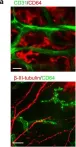
In Mucosal Immunol on 1 January 2020 by Wu, M., Liu, J., et al.
Fig.5.A

-
IHC-IF
-
Mus musculus (House mouse)
Collected and cropped from Mucosal Immunol by CiteAb, provided under a CC-BY license
Image 1 of 1