
Recent studies have revealed a structural role for DNA ligase 4 (Lig4) in the maintenance of a repair complex during non-homologous end joining (NHEJ) of DNA double-strand breaks. In cultured cell lines, catalytically inactive Lig4 can partially alleviate the severe DNA repair phenotypes observed in cells lacking Lig4. To study the structural role of Lig4 in vivo, a mouse strain harboring a point mutation to Lig4's catalytic site was generated. In contrast to the ablation of Lig4, catalytically inactive Lig4 mice are born alive. These mice display marked growth retardation and have clear deficits in lymphocyte development. We considered that the milder phenotype results from inactive Lig4 help to recruit another ligase to the repair complex. We next generated a mouse strain deficient for nuclear Lig3. Nuclear Lig3-deficient mice are moderately smaller and have elevated incidences of cerebral ventricle dilation but otherwise appear normal. Strikingly, in experiments crossing these two strains, mice lacking nuclear Lig3 and expressing inactive Lig4 were not obtained. Timed mating revealed that fetuses harboring both mutations underwent resorption, establishing an embryonic lethal genetic interaction. These data suggest that Lig3 is recruited to NHEJ complexes to facilitate end joining in the presence (but not activity) of Lig4.
© The Author(s) 2024. Published by Oxford University Press on behalf of Nucleic Acids Research.
Product Citations: 43
In Nucleic Acids Research on 11 January 2025 by Medina-Suárez, D., Han, L., et al.
-
Biochemistry and Molecular biology
-
Genetics
A TNIP1-driven systemic autoimmune disorder with elevated IgG4.
In Nature Immunology on 1 September 2024 by Medhavy, A., Athanasopoulos, V., et al.
Whole-exome sequencing of two unrelated kindreds with systemic autoimmune disease featuring antinuclear antibodies with IgG4 elevation uncovered an identical ultrarare heterozygous TNIP1Q333P variant segregating with disease. Mice with the orthologous Q346P variant developed antinuclear autoantibodies, salivary gland inflammation, elevated IgG2c, spontaneous germinal centers and expansion of age-associated B cells, plasma cells and follicular and extrafollicular helper T cells. B cell phenotypes were cell-autonomous and rescued by ablation of Toll-like receptor 7 (TLR7) or MyD88. The variant increased interferon-β without altering nuclear factor kappa-light-chain-enhancer of activated B cells signaling, and impaired MyD88 and IRAK1 recruitment to autophagosomes. Additionally, the Q333P variant impaired TNIP1 localization to damaged mitochondria and mitophagosome formation. Damaged mitochondria were abundant in the salivary epithelial cells of Tnip1Q346P mice. These findings suggest that TNIP1-mediated autoimmunity may be a consequence of increased TLR7 signaling due to impaired recruitment of downstream signaling molecules and damaged mitochondria to autophagosomes and may thus respond to TLR7-targeted therapeutics.
© 2024. The Author(s).
-
FC/FACS
-
Mus musculus (House mouse)
-
Immunology and Microbiology
In Cancer Cell on 8 April 2024 by Barišić, D., Chin, C. R., et al.
ARID1A, a subunit of the canonical BAF nucleosome remodeling complex, is commonly mutated in lymphomas. We show that ARID1A orchestrates B cell fate during the germinal center (GC) response, facilitating cooperative and sequential binding of PU.1 and NF-kB at crucial genes for cytokine and CD40 signaling. The absence of ARID1A tilts GC cell fate toward immature IgM+CD80-PD-L2- memory B cells, known for their potential to re-enter new GCs. When combined with BCL2 oncogene, ARID1A haploinsufficiency hastens the progression of aggressive follicular lymphomas (FLs) in mice. Patients with FL with ARID1A-inactivating mutations preferentially display an immature memory B cell-like state with increased transformation risk to aggressive disease. These observations offer mechanistic understanding into the emergence of both indolent and aggressive ARID1A-mutant lymphomas through the formation of immature memory-like clonal precursors. Lastly, we demonstrate that ARID1A mutation induces synthetic lethality to SMARCA2/4 inhibition, paving the way for potential precision therapy for high-risk patients.
Copyright © 2024 Elsevier Inc. All rights reserved.
-
Biochemistry and Molecular biology
-
Cancer Research
-
Immunology and Microbiology
-
Stem Cells and Developmental Biology
In JCI Insight on 8 February 2024 by Fuseya, Y., Kadoba, K., et al.
Linear ubiquitin chains, which are generated specifically by the linear ubiquitin assembly complex (LUBAC) ubiquitin ligase, play crucial roles in immune signaling, including NF-κB activation. LUBAC comprises catalytic large isoform of heme-oxidized iron regulatory protein 2 ubiquitin ligase 1 (HOIL-1L) interacting protein (HOIP), accessory HOIL-1L, and SHANK-associated RH domain-interacting protein (SHARPIN). Deletion of the ubiquitin ligase activity of HOIL-1L, an accessory ligase of LUBAC, augments LUBAC functions by enhancing LUBAC-mediated linear ubiquitination, which is catalyzed by HOIP. Here, we show that HOIL-1L ΔRING1 mice, which exhibit augmented LUBAC functions upon loss of the HOIL-1L ligase, developed systemic lupus erythematosus (SLE) and Sjögren's syndrome in a female-dominant fashion. Augmented LUBAC activity led to hyperactivation of both lymphoid and myeloid cells. In line with the findings in mice, we sought to identify missense single nucleotide polymorphisms/variations of the RBCK1/HOIL-1L gene in humans that attenuate HOIL-1L ligase activity. We found that the R464H variant, which is encoded by rs774507518 within the RBCK1/HOIL-1L gene, attenuated HOIL-1L ligase activity and augmented LUBAC-mediated immune signaling, including that mediated by Toll-like receptors. We also found that rs774507518 was enriched significantly in patients with SLE, strongly suggesting that RBCK1/HOIL-1L is an SLE susceptibility gene and that augmented linear ubiquitin signaling generated specifically by LUBAC underlies the pathogenesis of this prototype systemic autoimmune disease.
-
FC/FACS
-
Immunology and Microbiology
Antigen receptor signaling and cell death resistance controls intestinal humoral response zonation.
In Immunity on 10 October 2023 by Raso, F., Liu, S., et al.
Immunoglobulin A (IgA) maintains commensal communities in the intestine while preventing dysbiosis. IgA generated against intestinal microbes assures the simultaneous binding to multiple, diverse commensal-derived antigens. However, the exact mechanisms by which B cells mount broadly reactive IgA to the gut microbiome remains elusive. Here, we have shown that IgA B cell receptor (BCR) is required for B cell fitness during the germinal center (GC) reaction in Peyer's patches (PPs) and for generation of gut-homing plasma cells (PCs). We demonstrate that IgA BCR drove heightened intracellular signaling in mouse and human B cells, and as a consequence, IgA+ B cells received stronger positive selection cues. Mechanistically, IgA BCR signaling offset Fas-mediated death, possibly rescuing low-affinity B cells to promote a broad humoral response to commensals. Our findings reveal an additional mechanism linking BCR signaling, B cell fate, and antibody production location, which have implications for how intestinal antigen recognition shapes humoral immunity.
Copyright © 2023 Elsevier Inc. All rights reserved.
-
Immunology and Microbiology
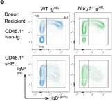
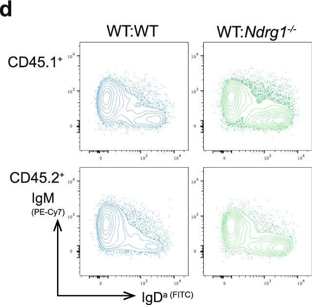
In Commun Biol on 10 November 2022 by Hodgson, R., Xu, X., et al.
Fig.4.E
-
FC/FACS
-
Mus musculus (House mouse)
Collected and cropped from Commun Biol by CiteAb, provided under a CC-BY license
Image 1 of 4
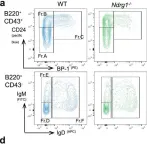
In Commun Biol on 10 November 2022 by Hodgson, R., Xu, X., et al.
Fig.3.A

-
FC/FACS
-
Mus musculus (House mouse)
Collected and cropped from Commun Biol by CiteAb, provided under a CC-BY license
Image 1 of 4
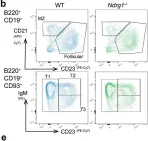
In Commun Biol on 10 November 2022 by Hodgson, R., Xu, X., et al.
Fig.3.B

-
FC/FACS
-
Mus musculus (House mouse)
Collected and cropped from Commun Biol by CiteAb, provided under a CC-BY license
Image 1 of 4
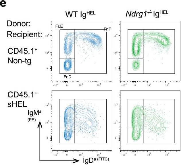
In Commun Biol on 10 November 2022 by Hodgson, R., Xu, X., et al.
Fig.5.D
-
FC/FACS
-
Mus musculus (House mouse)
Collected and cropped from Commun Biol by CiteAb, provided under a CC-BY license
Image 1 of 4