T follicular helper (TFH) cells are critical for humoral immunity during chronic viral infection, but the mechanisms guiding their differentiation from a novel CD4⁺ T cell progenitors remain incompletely understood. Themis, a T cell-specific adaptor protein, has been implicated in T cell development and function, but its role in peripheral CD4⁺ T cell differentiation under chronic antigen stimulation has not been defined.
We used a chronic lymphocytic choriomeningitis virus (LCMV) Clone13 infection model in wild-type and Themis conditional knockout (cKO) mice. A combination of adoptive cell transfer, flow cytometry, histological analysis, and single-cell RNA sequencing (scRNA-seq) was applied to analyze the differentiation of CD4⁺ T cells into TFH cells at multiple infection stages.
Themis expression is strongly upregulated in TFH cells at early stages of infection, and as expected, Themis promotes TFH cell differentiation at this stage. However, unexpectedly, at the late stages of chronic LCMV infection, Themis-deficient CD4+ T cells favored TFH cell differentiation and helped control the virus by enhancing GC responses and antibody production, suggesting that Themis inhibits TFH cell differentiation at this stage. In the late stage we found that Themis inhibits the differentiation of CD4+ T cell progenitors into TFH cells through transcriptional regulation.
Our study uncovers a dual-stage regulatory role of Themis in TFH cell differentiation during chronic viral infection. While promoting TFH generation early, Themis unexpectedly restrains excessive differentiation at later stages, suggesting its function is context- and time-dependent. These findings highlight Themis as a key temporal regulator of CD4⁺ T cell fate decisions under chronic antigenic stress.
Copyright © 2025 Zhu, Bao, Wang, Gan, Tang, Cong, Hou, Quan, Yan, Liu, Lin, Zhang, Du, Hou, Gascoigne, Xu, Fu and Zheng.
Product Citations: 17
In Frontiers in Immunology on 8 August 2025 by Zhu, Y., Bao, Y., et al.
-
Immunology and Microbiology
In Molecular Therapy on 3 July 2024 by Cheang, N. Y. Z., Tan, K. S., et al.
Current coronavirus disease 2019 vaccines face limitations including waning immunity, immune escape by severe acute respiratory syndrome coronavirus 2 (SARS-CoV-2) variants, limited cellular response, and poor mucosal immunity. We engineered a Clec9A-receptor binding domain (RBD) antibody construct that delivers the SARS-CoV-2 RBD to conventional type 1 dendritic cells. Compared with non-targeting approaches, single dose immunization in mice with Clec9A-RBD induced far higher RBD-specific antibody titers that were sustained for up to 21 months after vaccination. Uniquely, increasing neutralizing and antibody-dependent cytotoxicity activities across the sarbecovirus family was observed, suggesting antibody affinity maturation over time. Consistently and remarkably, RBD-specific follicular T helper cells and germinal center B cells persisted up to 12 months after immunization. Furthermore, Clec9A-RBD immunization induced a durable mono- and poly-functional T-helper 1-biased cellular response that was strongly cross-reactive against SARS-CoV-2 variants of concern, including Omicron subvariants, and with a robust CD8+ T cell signature. Uniquely, Clec9A-RBD single-shot systemic immunization effectively primed RBD-specific cellular and humoral immunity in lung and resulted in significant protection against homologous SARS-CoV-2 challenge as evidenced by limited body weight loss and approximately 2 log10 decrease in lung viral loads compared with non-immunized controls. Therefore, Clec9A-RBD immunization has the potential to trigger robust and sustained, systemic and mucosal protective immunity against rapidly evolving SARS-CoV2 variants.
Copyright © 2024 The Author(s). Published by Elsevier Inc. All rights reserved.
-
COVID-19
-
Immunology and Microbiology
In Frontiers in Immunology on 17 September 2021 by Yang, L., Chen, W., et al.
Follicular helper T (TFH) cells are specialized CD4+ helper T cells that provide help to B cells in humoral immunity. However, the molecular mechanism underlying generation of TFH cells is incompletely understood. Here, we reported that Damage-specific DNA binding protein 1 (Ddb1) was required for expansion of CD4+ helper T cells including TFH and Th1 cells, germinal center response, and antibody response to acute viral infection. Ddb1 deficiency in activated CD4+ T cells resulted in cell cycle arrest at G2-M phase and increased cell death, due to accumulation of DNA damage and hyperactivation of ATM/ATR-Chk1 signaling. Moreover, mice with deletion of both Cul4a and Cul4b in activated CD4+ T cells phenocopied Ddb1-deficient mice, suggesting that E3 ligase-dependent function of Ddb1 was crucial for genome maintenance and helper T-cell generation. Therefore, our results indicate that Ddb1 is an essential positive regulator in the expansion of CD4+ helper T cells.
Copyright © 2021 Yang, Chen, Li, Xiao, Fan, Zhang, Xia, Li, Hong, Zhao, Li, Liu and Xiao.
-
Immunology and Microbiology
Endogenous retroviruses promote homeostatic and inflammatory responses to the microbiota.
In Cell on 8 July 2021 by Lima-Júnior, D. S., Krishnamurthy, S. R., et al.
The microbiota plays a fundamental role in regulating host immunity. However, the processes involved in the initiation and regulation of immunity to the microbiota remain largely unknown. Here, we show that the skin microbiota promotes the discrete expression of defined endogenous retroviruses (ERVs). Keratinocyte-intrinsic responses to ERVs depended on cyclic GMP-AMP synthase (cGAS)/stimulator of interferon genes protein (STING) signaling and promoted the induction of commensal-specific T cells. Inhibition of ERV reverse transcription significantly impacted these responses, resulting in impaired immunity to the microbiota and its associated tissue repair function. Conversely, a lipid-enriched diet primed the skin for heightened ERV- expression in response to commensal colonization, leading to increased immune responses and tissue inflammation. Together, our results support the idea that the host may have co-opted its endogenous virome as a means to communicate with the exogenous microbiota, resulting in a multi-kingdom dialog that controls both tissue homeostasis and inflammation.Published by Elsevier Inc.
-
Immunology and Microbiology
BNT162b vaccines protect rhesus macaques from SARS-CoV-2.
In Nature on 1 April 2021 by Vogel, A. B., Kanevsky, I., et al.
A safe and effective vaccine against COVID-19 is urgently needed in quantities that are sufficient to immunize large populations. Here we report the preclinical development of two vaccine candidates (BNT162b1 and BNT162b2) that contain nucleoside-modified messenger RNA that encodes immunogens derived from the spike glycoprotein (S) of SARS-CoV-2, formulated in lipid nanoparticles. BNT162b1 encodes a soluble, secreted trimerized receptor-binding domain (known as the RBD-foldon). BNT162b2 encodes the full-length transmembrane S glycoprotein, locked in its prefusion conformation by the substitution of two residues with proline (S(K986P/V987P); hereafter, S(P2) (also known as P2 S)). The flexibly tethered RBDs of the RBD-foldon bind to human ACE2 with high avidity. Approximately 20% of the S(P2) trimers are in the two-RBD 'down', one-RBD 'up' state. In mice, one intramuscular dose of either candidate vaccine elicits a dose-dependent antibody response with high virus-entry inhibition titres and strong T-helper-1 CD4+ and IFNγ+CD8+ T cell responses. Prime-boost vaccination of rhesus macaques (Macaca mulatta) with the BNT162b candidates elicits SARS-CoV-2-neutralizing geometric mean titres that are 8.2-18.2× that of a panel of SARS-CoV-2-convalescent human sera. The vaccine candidates protect macaques against challenge with SARS-CoV-2; in particular, BNT162b2 protects the lower respiratory tract against the presence of viral RNA and shows no evidence of disease enhancement. Both candidates are being evaluated in phase I trials in Germany and the USA1-3, and BNT162b2 is being evaluated in an ongoing global phase II/III trial (NCT04380701 and NCT04368728).
-
COVID-19
-
Immunology and Microbiology
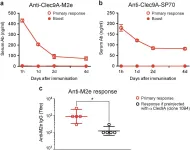
In NPJ Vaccines on 22 December 2017 by Park, H. Y., Tan, P. S., et al.
Fig.3.A,B,C

-
ELISA
-
Mus musculus (House mouse)
Collected and cropped from NPJ Vaccines by CiteAb, provided under a CC-BY license
Image 1 of 1