Attenuated whole organism vaccines targeting the malaria liver stage reliably confer sterile immunity. These vaccines completely protect female mice from infection, but protection in male mice remains unproven. We discover that male mice vaccinated with prime-and-trap, a whole organism-based vaccine strategy, exhibit poorer protection against Plasmodium sporozoite challenge than females. We investigate this sex difference, and identify vaccinated males have fewer hepatic memory CD8+ T cells than females when scaling for liver biomass, and reduced inflammatory responses post-vaccination. Surgical hormone manipulation clarifies that the presence of testicular hormones hinders protection in male mice. The presence of androgens does not affect memory CD8+ T cell quantity nor quality, but reduces recruitment of CD8+ T cells in male liver tissues via a restricted inflammatory response. Here, we show both males and females form functional memory responses following prime-and-trap vaccination, but the presence of androgens during sporozoite challenge impair protection in male mice.
© 2025. The Author(s).
Product Citations: 63
In Nature Communications on 4 June 2025 by Duncombe, C. J., Sen, N., et al.
-
FC/FACS
-
Mus musculus (House mouse)
-
Immunology and Microbiology
B cells modulate lung antiviral inflammatory responses via the neurotransmitter acetylcholine.
In Nature Immunology on 1 May 2025 by Cembellin-Prieto, A., Luo, Z., et al.
The rapid onset of innate immune defenses is critical for early control of viral replication in an infected host and yet it can also lead to irreversible tissue damage, especially in the respiratory tract. Sensitive regulators must exist that modulate inflammation, while controlling the infection. In the present study, we identified acetylcholine (ACh)-producing B cells as such early regulators. B cells are the most prevalent ACh-producing leukocyte population in the respiratory tract demonstrated with choline acetyltransferase (ChAT)-green fluorescent protein (GFP) reporter mice, both before and after infection with influenza A virus. Mice lacking ChAT in B cells, disabling their ability to generate ACh (ChatBKO), but not those lacking ChAT in T cells, significantly, selectively and directly suppressed α7-nicotinic-ACh receptor-expressing interstitial, but not alveolar, macrophage activation and their ability to secrete tumor necrosis factor (TNF), while better controlling virus replication at 1 d postinfection. Conversely, TNF blockade via monoclonal antibody treatment increased viral loads at that time. By day 10 of infection, ChatBKO mice showed increased local and systemic inflammation and reduced signs of lung epithelial repair despite similar viral loads and viral clearance. Thus, B cells are key participants of an immediate early regulatory cascade that controls lung tissue damage after viral infection, shifting the balance toward reduced inflammation at the cost of enhanced early viral replication.
© 2025. The Author(s).
-
Immunology and Microbiology
In Vaccines on 1 March 2025 by Lu, B., Chaudhary, O., et al.
Although mRNA vaccines have the potential to be developed and deployed rapidly to combat infectious diseases, the ideal method of administration and boosting schedule strategy for generating optimal immunogenicity is an area of active research. We compared the immune responses resulting from different schedules for prime-boost and boosting either ipsilaterally or contralaterally in relation to the initial vaccine dose.
Influenza hemagglutinin (HA) was used as a model antigen for different vaccination regimens in mice using both mRNA lipid nanoparticles (mRNA-LNP) and AF03-adjuvanted recombinant protein (rHA-AF03) vaccines.
Increasing the prime-boost interval resulted in higher levels of serum anti-HA IgG and functional antibody hemagglutination inhibition (HAI) responses in mRNA-LNP-vaccinated animals, which correlated with an induction of germinal center (GC) B cells and follicular helper T (Tfh) cells in lymph nodes. In addition, longer prime-boost intervals resulted in higher levels of IL-2 and TNF-α producing CD4+ T cells two weeks after boosting. The number of Ig-secreting long-lived plasma cells increased with the length of prime-boost intervals. Contralateral boosting resulted in an increase in HAI titers and GC B cells compared to an ipsilateral boost. However, significantly higher numbers of GC B cells were induced in the draining lymph nodes following ipsilateral boosting than in the non-draining lymph nodes.
Overall, our data provides insights into the immune mechanisms of action of mRNA-LNP to develop the optimal vaccine regimen for mRNA vaccine platforms.
-
Genetics
-
Immunology and Microbiology
In Cell Reports Medicine on 16 April 2024 by Lim, R. J., Salehi-Rad, R., et al.
Immune checkpoint blockade (ICB) with PD-1/PD-L1 inhibition has revolutionized the treatment of non-small cell lung cancer (NSCLC). Durable responses, however, are observed only in a subpopulation of patients. Defective antigen presentation and an immunosuppressive tumor microenvironment (TME) can lead to deficient T cell recruitment and ICB resistance. We evaluate intratumoral (IT) vaccination with CXCL9- and CXCL10-engineered dendritic cells (CXCL9/10-DC) as a strategy to overcome resistance. IT CXCL9/10-DC leads to enhanced T cell infiltration and activation in the TME and tumor inhibition in murine NSCLC models. The antitumor efficacy of IT CXCL9/10-DC is dependent on CD4+ and CD8+ T cells, as well as CXCR3-dependent T cell trafficking from the lymph node. IT CXCL9/10-DC, in combination with ICB, overcomes resistance and establishes systemic tumor-specific immunity in murine models. These studies provide a mechanistic understanding of CXCL9/10-DC-mediated host immune activation and support clinical translation of IT CXCL9/10-DC to augment ICB efficacy in NSCLC.
Published by Elsevier Inc.
-
Mus musculus (House mouse)
-
Cancer Research
-
Immunology and Microbiology
In Communications Biology on 20 April 2023 by Zhang, Y., Do, K. K., et al.
The cornea is the outmost ocular tissue and plays an important role in protecting the eye from environmental insults. Corneal epithelial wounding provokes pain and fear and contributes to the most ocular trauma emergency assessments worldwide. ZEB1 is an essential transcription factor in development; but its roles in adult tissues are not clear. We identify Zeb1 is an intrinsic factor that facilitates corneal epithelial wound healing. In this study, we demonstrate that monoallelic deletion of Zeb1 significantly expedites corneal cell death and inhibits corneal epithelial EMT-related cell migration upon an epithelial debridement. We provide evidence that Zeb1-regulation of corneal epithelial wound healing is through the repression of genes required for Tnfa-induced epithelial cell death and the induction of genes beneficial for epithelial cell migration. We suggest utilizing TNF-α antagonists would reduce TNF/TNFR1-induced cell death in the corneal epithelium and inflammation in the corneal stroma to help corneal wound healing.
© 2023. The Author(s).
-
IHC
-
Mus musculus (House mouse)
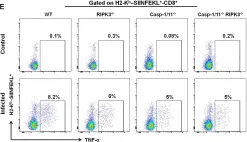
In Front Immunol on 25 April 2020 by Rana, A., de Almeida, F. C., et al.
Fig.5.E

-
FC/FACS
-
Mus musculus (House mouse)
Collected and cropped from Front Immunol by CiteAb, provided under a CC-BY license
Image 1 of 1