Variant-adapted vaccines are recommended in vulnerable populations to address the waning immunity and the emergence of immune-escaping SARS-CoV-2 variants, yet data about immune responses to such vaccines in people living with HIV (PLWH) are limited. We therefore aimed to assess long-term immune responses to an original-BA.4/5 mRNA booster in this population.
In this prospective longitudinal study, PLWH receiving either an original-BA.4/5 bivalent booster or an original monovalent booster and HIV-negative healthcare workers (HCWs) receiving a bivalent booster were enrolled and sampled before (T0), 1 month (T1), and 4-9 months (T2) after the vaccine administration. SARS-CoV-2-specific T and B cells, RBD-binding antibodies, and RBD-blocking antibodies against both wild type (WT) and omicron BA.4/5 virus were determined.
The bivalent booster is able to transiently increase both humoral and polyfunctional T cell responses in PLWH, with humoral responses comparable to those observed in HCWs. While T cell responses are cross-reactive against viral variants and stable over time, humoral immunity is imprinted to the ancestral virus and wanes quickly. Furthermore, whilst previous SARS-CoV-2 infection does not affect the trajectory of vaccine-elicited immune responses, markers of HIV-related T cell dysfunction are associated with lower antibody peak responses and higher antibody waning. Lastly, the bivalent booster was superior to the monovalent one in inducing BA.4/5-reactive RBD-blocking antibodies.
The original-BA.4/5 bivalent booster is highly immunogenic in PLWH and superior to the monovalent one in inducing humoral responses against the BA.4/5 virus, although HIV-related T cell dysfunction markers are associated with blunted and less durable antibody immunity.
© 2025. The Author(s).
Product Citations: 135
Long-term immune responses to SARS-CoV-2 Omicron BA.4/5 mRNA booster in people living with HIV.
In Commun Med (Lond) on 27 March 2025 by Augello, M., Bono, V., et al.
-
SARS Coronavirus (SARS-CoV)
-
COVID-19
-
Genetics
-
Immunology and Microbiology
Decrease of NAD+ Inhibits the Apoptosis of OLP T Cells via Inducing Mitochondrial Fission.
In Journal of Inflammation Research on 28 January 2025 by Zhang, Z. Y., Wang, F., et al.
Oral lichen planus (OLP) is a chronic, immune-mediated inflammatory disease involving T cells. Mitochondrial fission plays a crucial role in T cell fate through structural remodeling. Nicotinamide adenine dinucleotide (NAD+) regulates mitochondrial remodeling and function. This study explored the role of NAD+ in modulating mitochondrial fission and apoptosis in T cells under the OLP immune-inflammatory environment.
T cells and plasma were isolated from peripheral blood. Mitochondrial morphology was characterized by transmission electron microscopy and Mito-Tracker staining. OLP plasma-exposed Jurkat T cells were infected with the Drp1 shRNA virus to investigate the role of mitochondrial fission in OLP T cell apoptosis. OLP T cells and OLP plasma-exposed Jurkat T cells were treated with either β-nicotinamide mononucleotide (an NAD+ synthesis precursor) or FK866 (an NAD+ synthesis inhibitor) to assess the effect of NAD+ regulation on mitochondrial remodeling and T cell apoptosis.
OLP T cells exhibited fragmented mitochondria with elevated dynamin-related protein 1 (Drp1) and reduced mitofusin 2 (Mfn2) expression, accompanied by decreased apoptosis. Drp1 knockdown in OLP plasma-exposed Jurkat T cells increased apoptosis and reduced proliferation. NAD+ levels were reduced in both OLP T cells and OLP plasma-treated Jurkat T cells, leading to enhanced mitochondrial fission, decreased mitochondrial membrane potential (MMP) and respiration function, and reduced apoptosis rate. β-nicotinamide mononucleotide supplementation restored NAD+ levels, suppressed mitochondrial fission, improved MMP, and promoted apoptosis in these cells.
Reduced NAD+ levels in OLP T cells enhanced mitochondrial fission and contributed to decreased apoptosis. NAD+ supplementation mitigated these effects, suggesting a potential therapeutic strategy for restoring T cell homeostasis in OLP.
© 2025 Zhang et al.
-
Homo sapiens (Human)
-
Cell Biology
-
Immunology and Microbiology
In Front Aging on 9 September 2024 by Frasca, D. & Bueno, V.
In this paper, we measured B cell function in elderly healthy individuals (EH) and in elderly patients with Type-2 Diabetes Mellitus (T2DM, ET2DM), which are treatment-naive, as compared to healthy young (YH) individuals. Results show a higher serum inflammatory status of elderly versus young individuals, and especially of ET2DM versus EH. This status is associated with a reduced response to the seasonal influenza vaccine and with increased frequencies of the circulating pro-inflammatory B cell subset called Double Negative (DN) B cells. B cells from ET2DM patients are not only more inflammatory but also hyper-metabolic as compared to those from EH controls. The results herein are to our knowledge the first to show that T2DM superimposed on aging further increases systemic and B cell intrinsic inflammation, as well as dysfunctional humoral immunity. Our findings confirm and extend our previously published findings showing that inflammatory B cells are metabolically supported.
Copyright © 2024 Frasca and Bueno.
-
FC/FACS
-
Cell Biology
-
Immunology and Microbiology
Conserved epigenetic hallmarks of T cell aging during immunity and malignancy.
In Nature Aging on 1 August 2024 by Mi, T., Soerens, A. G., et al.
Chronological aging correlates with epigenetic modifications at specific loci, calibrated to species lifespan. Such 'epigenetic clocks' appear conserved among mammals, but whether they are cell autonomous and restricted by maximal organismal lifespan remains unknown. We used a multilifetime murine model of repeat vaccination and memory T cell transplantation to test whether epigenetic aging tracks with cellular replication and if such clocks continue 'counting' beyond species lifespan. Here we found that memory T cell epigenetic clocks tick independently of host age and continue through four lifetimes. Instead of recording chronological time, T cells recorded proliferative experience through modification of cell cycle regulatory genes. Applying this epigenetic profile across a range of human T cell contexts, we found that naive T cells appeared 'young' regardless of organism age, while in pediatric patients, T cell acute lymphoblastic leukemia appeared to have epigenetically aged for up to 200 years. Thus, T cell epigenetic clocks measure replicative history and can continue to accumulate well-beyond organismal lifespan.
© 2024. The Author(s).
-
FC/FACS
-
Homo sapiens (Human)
-
Genetics
-
Immunology and Microbiology
Reconstitution of human microglia and resident T cells in the brain of humanized DRAGA mice.
In Frontiers in Cellular and Infection Microbiology on 10 July 2024 by Ghosh Roy, S., Karim, A. F., et al.
Humanized mouse models are valuable tools for investigating the human immune system in response to infection and injury. We have previously described the human immune system (HIS)-DRAGA mice (HLA-A2.HLA-DR4.Rag1KO.IL-2RgKO.NOD) generated by infusion of Human Leukocyte Antigen (HLA)-matched, human hematopoietic stem cells from umbilical cord blood. By reconstituting human cells, the HIS-DRAGA mouse model has been utilized as a "surrogate in vivo human model" for infectious diseases such as Human Immunodeficiency Virus (HIV), Influenza, Coronavirus Disease 2019 (COVID-19), scrub typhus, and malaria. This humanized mouse model bypasses ethical concerns about the use of fetal tissues for the humanization of laboratory animals. Here in, we demonstrate the presence of human microglia and T cells in the brain of HIS-DRAGA mice. Microglia are brain-resident macrophages that play pivotal roles against pathogens and cerebral damage, whereas the brain-resident T cells provide surveillance and defense against infections. Our findings suggest that the HIS-DRAGA mouse model offers unique advantages for studying the functions of human microglia and T cells in the brain during infections, degenerative disorders, tumors, and trauma, as well as for testing therapeutics in these pathological conditions.
Copyright © 2024 Ghosh Roy, Karim, Brumeanu and Casares.
-
Immunology and Microbiology
-
Neuroscience
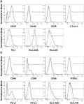
In Front Immunol on 29 January 2019 by Wefers, C., Duiveman-de Boer, T., et al.
Fig.4.A

-
FC/FACS
-
Homo sapiens (Human)
Collected and cropped from Front Immunol by CiteAb, provided under a CC-BY license
Image 1 of 2
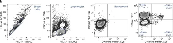
In PLoS One on 15 December 2015 by Vir, P., Arrigucci, R., et al.
Fig.1.B

-
FC/FACS
-
Collected and cropped from PLoS One by CiteAb, provided under a CC-BY license
Image 1 of 2