Mesenchymal stem cells (MSCs) are frequently used for therapeutic applications in both pre-clinical and clinical settings owing to their capacity for immune modulation and neuroprotective effects. However, transient fever is commonly observed as an adverse event following MSC injection in patients with Alzheimer's disease (AD). In this study, we investigated the potential impact of immunosuppressants such as dexamethasone and tacrolimus on altering the characteristics of human mesenchymal stem cells (hMSCs). Additionally, we examined whether these immunosuppressants affect the persistence of hMSCs or the immune response upon their administration into the brain parenchyma of AD mice. The exposure of hMSCs to high concentrations of dexamethasone and tacrolimus in vitro did not significantly alter the characteristics of hMSCs. The expression of genes related to innate immune responses, such as Irak1, Irf3, Nod1, and Ifnar1, was significantly downregulated by the additional administration of dexamethasone and tacrolimus to the brain parenchyma of AD mice. However, hMSC persistence in the AD mouse brain was not affected. The results of this study support the use of immunosuppressants to mitigate fever during stem cell therapy in patients with AD.
Product Citations: 183
In International Journal of Stem Cells on 30 May 2025 by Lee, N. K., Na, D. L., et al.
-
Stem Cells and Developmental Biology
In Clinical and Experimental Immunology on 21 January 2025 by Zagrodnik, J. L., Blandford, S. N., et al.
Multiple sclerosis (MS) is a chronic immune-mediated demyelinating disease of the central nervous system, whereby clinical disease activity is primarily monitored by magnetic resonance imaging.
Given the limitations associated with implementing and acquiring novel and emerging imaging biomarkers in routine clinical practice, the discovery of biofluid biomarkers may offer a more simple and cost-effective measure that would improve accessibility, standardization, and patient care. Extracellular vesicles (EVs) are nanoparticles secreted from cells under both homeostatic and pathological states, and have been recently investigated as biomarkers in MS. The objectives of this study were to longitudinally measure levels of specific immune cell-derived EVs in MS and provide evidence that EV sub-populations may serve as biomarkers of disease activity, axonal injury, and/or clinical disability.
Our results demonstrate that the rate of clinical disability in MS negatively correlates with changes in circulating CD3+ EVs within the plasma. Additionally, numbers of CD4+ EVs decrease in individuals with increasing pNfL levels overtime whereby the magnitude of the pNfL increase negatively correlates with changes in plasma CD4+ and CD8+ EVs. Finally, when applying NEDA-3 criteria to define active versus stable disease, individuals with active disease had significantly elevated CD4+ and CD8+ EVs compared to stable disease.
In summary, the analysis of specific immune cell-derived EV subsets may provide a method to monitor disability accumulation, disease activity, and axonal injury in MS, while also providing insights into the pathophysiology and cellular/molecular mechanisms that influence progression.
© The Author(s) 2025. Published by Oxford University Press on behalf of the British Society for Immunology.
-
Immunology and Microbiology
Preprint on BioRxiv : the Preprint Server for Biology on 19 January 2025 by Escós, A., Ambikan, A. T., et al.
Cells of the myeloid lineage, particularly monocytes and macrophages, play a key role in HIV infection by contributing to viral replication, immune response, and maintaining immune balance during suppressive therapy. We hypothesized that metabolic reprogramming and altered chemokine signaling in people living with HIV (PWH) on long-term antiretroviral therapy (ART) affect monocyte transport and polarization due to ongoing inflammation. Therefore, the present study aimed to identify the mechanism of impaired monocyte/macrophage function in PWH on well-treated ART that can lead to clinical intervention strategies to improve health. Single-cell RNA sequencing, immune-phenotyping, and metabolic modeling identified altered expression of chemokine and metabolite receptors and altered metabolic flux in PWH monocytes that decreased monocyte migration. The plasma secretome revealed a nonclassical inflammatory microenvironment in PWH. Integrative multi-omics and single-cell proteomics of differentiated monocyte-derived macrophages (MDMs) detected metabolic reprogramming orchestrated by α-ketoglutarate (AKG) that affected macrophage function and HIV infection. Increased levels of AKG in plasma were shown to occur in PWH under ART. Therefore, when differentiating MDM with serum from PWH or AKG, macrophage function was found polarized towards an M2-like state. AKG alone was shown to increase CCR5 levels and increase HIV-1 infection in MDM. Here, we utilize systems biology-driven identification and ex vivo assays to show impaired macrophage polarization, due to metabolic training, can leads to a low-grade nonclassical inflammatory environment in well-treated PWH.
-
FC/FACS
-
Immunology and Microbiology
In Veterinary Research on 18 December 2024 by Huang, N., Ye, L., et al.
Diarrhoea and preweaning mortality in piglets are crucial factors impacting the economic sustainability of the swine industry. Pathogenic infections are among the main causes of diarrhea and mortality. Group 3 innate lymphoid cells (ILC3s) are crucial for safeguarding against pathogenic infections. However, knowledge regarding the development and function of ILC3s in suckling piglets is currently limited. Our findings demonstrate that the development of ILC3s in suckling piglets gradually progresses from day 1 to day 21, with a notable increase observed on day 28. Additionally, the development of NKp46+ILC3s and the production of interleukin (IL)-17A by ILC3s displayed consistent patterns with the changes observed in ILC3s. Notably, interferon (IFN)-γ levels significantly increased on day 14. Moreover, the production of IFN-γ by NKp46+ILC3s was greater than that by NKp46-ILC3s. Importantly, when piglets were subjected to a 4-h challenge with enterotoxigenic Escherichia coli, both the percentages of ILC3s significantly increased, accompanied by increased IL-22 production, highlighting their importance in maintaining intestinal health. The outcomes of this study provide valuable insights for future related research.
© 2024. The Author(s).
-
Immunology and Microbiology
-
Stem Cells and Developmental Biology
-
Veterinary Research
In Clinical Interventions in Aging on 27 November 2024 by Liu, D. & Chi, L. J.
Vascular cognitive impairment(VCI) ranks as the second most prevalent type of dementia.Increasing evidence has shown that inflammation and multi-faceted neuro-immune interactions integrate systemic and central inflammatory pathways, thereby inducing vascular tissue injury and contributing to the development of vascular cognitive impairment (VCI).V-type immunoglobulin-like suppressor of T cell activation (VISTA) is an Negative checkpoint regulators(NCR) that is associated with CNS homeostasis, interactions with peripheral immunity and CNS inflammation.The primary objective of this study was to seek the correlation between VISTA and VCI in patients with cardiovascular risk factors.Our secondary objective was to explore the potential of VISTA as a biomarker for VCI.
We enrolled individuals with cardiovascular risk factors in this cross-sectional study research and categorized them into two groups: without cognitive impairment (control) and with cognitive impairment (VCI). VISTA expression in peripheral blood mononuclear cells (PBMCs) was analyzed using relative quantitative polymerase chain reaction. VISTA expression was identified in monocyte subsets using flow cytometry. We use Enzyme linked immunosorbent assay to detect inflammatory factors in serum.
In PBMC in patients with VCI, the expression of VSIR was significantly reduced. In contrast to controls, fasting glucose, fibrosis, and the levels of interleukin 6 (IL-6) in VCI patients were noticeably higher, and uric acid levels were significantly lower. Vsir mRNA expression in PBMCs correlated negatively with IL-6 levels, Trail Making Test B scores, and Hachinski scores and positively with Boston Naming Test scores. In intermediate monocytes, flow cytometry showed reduced Vsir expression, which was connected with VCI. The percentage of intermediate monocytes, uric acid, and the VISTA mean fluorescence intensity on intermediate monocytes were shown to be independent factors to VCI by multivariate logistic regression analysis.
Decreased VISTA promotes the occurrence of VCI in patients with cardiovascular risk factors by promoting monocytes toward the proinflammatory intermediate monocyte subset. VISTA may serve as a potential biomarker for distinguishing VCI in individuals with cardiovascular risk factors.
© 2024 Liu and Chi.
-
Homo sapiens (Human)
-
Neuroscience
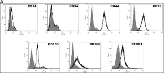
In PLoS One on 30 August 2014 by Tsai, H. L., Deng, W. P., et al.
Fig.1.A

-
WB
-
Homo sapiens (Human)
Collected and cropped from PLoS One by CiteAb, provided under a CC-BY license
Image 1 of 2
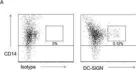
In PLoS Pathog on 1 January 2013 by Chen, Y., Hwang, S. L., et al.
Fig.5.A

-
FC/FACS
-
Collected and cropped from PLoS Pathog by CiteAb, provided under a CC-BY license
Image 1 of 2