The identification of affordable and easily accessible indicators to predict overall survival is important for tumor immunotherapy. Myeloid-derived suppressor cells (MDSCs) are a heterogeneous population of immature myeloid cells, which promote tumor immune escape in the tumor microenvironment (TME). This study aimed to determine whether peripheral blood MDSCs could determine their potential as predictors of survival in tumor patients with immunotherapy.
Flow cytometry was used to detect peripheral blood monocytic myeloid-derived suppressor cells (M-MDSCs) and granulocytic myeloid-derived suppressor cells (G-MDSCs) in 126 patients. Multivariate Cox regression analysis was conducted to examine the associations between peripheral blood MDSCs and patient survival. The receiver operating characteristic (ROC) curve determined the optimal cutoff value for peripheral blood MDSCs and grouped the indicators. The relationship between peripheral blood M-MDSCs and the prognosis and treatment outcome of tumor patients was explored.
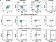
The proportion of peripheral blood M-MDSCs was associated with the prognosis of patients with tumors, as were tumor metastasis, the red blood cell count, absolute neutrophil count, absolute monocyte count, and BMI. Multivariate Cox regression analysis revealed that M-MDSCs, absolute lymphocyte value, and tumor metastasis were independent risk factors affecting the prognosis of patients with tumors. Detection of peripheral blood M-MDSCs obtained high sensitivity and specificity for tumor diagnosis. Patients with high M-MDSCs percentage demonstrated reduced survival durations and diminished responses to immunotherapy compared to those with low M-MDSCs percentage.
Peripheral blood M-MDSCs may be used to predict overall survival and immunotherapy efficacy outcomes. This study provides a putative predictive biomarker for clinicians to choose from to predict tumor patients' survival and the selection of receiving immunotherapy regimens.
© 2025. The Author(s).
Product Citations: 39
In BMC Immunology on 24 May 2025 by Sheng, W., Ding, Y., et al.
-
FC/FACS
-
Homo sapiens (Human)
-
Cancer Research
-
Cardiovascular biology
-
Immunology and Microbiology
Immune perturbations in human pancreas lymphatic tissues prior to and after type 1 diabetes onset.
In Nature Communications on 18 May 2025 by Golden, G. J., Wu, V. H., et al.
Autoimmune destruction of pancreatic β cells results in type 1 diabetes (T1D), with pancreatic immune infiltrate representing a key feature in this process. However, characterization of the immunological processes occurring in human pancreatic lymphatic tissues is lacking. Here, we conduct a comprehensive study of immune cells from pancreatic, mesenteric, and splenic lymphatic tissues of non-diabetic control (ND), β cell autoantibody-positive non-diabetic (AAb+), and T1D donors using flow cytometry and CITEseq. Compared to ND pancreas-draining lymph nodes (pLN), AAb+ and T1D donor pLNs display decreased CD4+ Treg and increased stem-like CD8+ T cell signatures, while only T1D donor pLNs exhibit naive T cell and NK cell differentiation. Mesenteric LNs have modulations only in CD4+ Tregs and naive cells, while splenocytes lack these perturbations. Further, T cell expression of activation markers and IL7 receptor correlate with T1D genetic risk. These results demonstrate tissue-restricted immune changes occur before and after T1D onset.
© 2025. The Author(s).
-
FC/FACS
-
Homo sapiens (Human)
-
Immunology and Microbiology
In STAR Protocols on 20 September 2024 by Li, Z., Ma, R., et al.
Type 2 innate lymphoid cells (ILC2s) are crucial in regulating immune responses and various physiological processes, including tissue repair, metabolic homeostasis, inflammation, and cancer surveillance. Here, we present a protocol that outlines the isolation, expansion, and adoptive transfer of human ILC2s from peripheral blood mononuclear cells for an in vivo lineage tracking experiment in a mouse model. Additionally, we detail the steps involved in the adoptive transfer of human ILC2s to recipient mice bearing human liquid or solid tumors. For complete details on the use and execution of this protocol, please refer to Li et al.1.
Copyright © 2024. Published by Elsevier Inc.
-
Homo sapiens (Human)
-
Cancer Research
N-MYC regulates cell survival via eIF4G1 in inv(16) acute myeloid leukemia.
In Science Advances on 1 March 2024 by Peramangalam, P. S., Surapally, S., et al.
N-MYC (encoded by MYCN) is a critical regulator of hematopoietic stem cell function. While the role of N-MYC deregulation is well established in neuroblastoma, the importance of N-MYC deregulation in leukemogenesis remains elusive. Here, we demonstrate that N-MYC is overexpressed in acute myeloid leukemia (AML) cells with chromosome inversion inv(16) and contributes to the survival and maintenance of inv(16) leukemia. We identified a previously unknown MYCN enhancer, active in multiple AML subtypes, essential for MYCN mRNA levels and survival in inv(16) AML cells. We also identified eukaryotic translation initiation factor 4 gamma 1 (eIF4G1) as a key N-MYC target that sustains leukemic survival in inv(16) AML cells. The oncogenic role of eIF4G1 in AML has not been reported before. Our results reveal a mechanism whereby N-MYC drives a leukemic transcriptional program and provides a rationale for the therapeutic targeting of the N-MYC/eIF4G1 axis in myeloid leukemia.
-
Cancer Research
In Cell on 1 February 2024 by Li, Z., Ma, R., et al.
The therapeutic potential for human type 2 innate lymphoid cells (ILC2s) has been underexplored. Although not observed in mouse ILC2s, we found that human ILC2s secrete granzyme B (GZMB) and directly lyse tumor cells by inducing pyroptosis and/or apoptosis, which is governed by a DNAM-1-CD112/CD155 interaction that inactivates the negative regulator FOXO1. Over time, the high surface density expression of CD155 in acute myeloid leukemia cells impairs the expression of DNAM-1 and GZMB, thus allowing for immune evasion. We describe a reliable platform capable of up to 2,000-fold expansion of human ILC2s within 4 weeks, whose molecular and cellular ILC2 profiles were validated by single-cell RNA sequencing. In both leukemia and solid tumor models, exogenously administered expanded human ILC2s show significant antitumor effects in vivo. Collectively, we demonstrate previously unreported properties of human ILC2s and identify this innate immune cell subset as a member of the cytolytic immune effector cell family.
Copyright © 2023 Elsevier Inc. All rights reserved.
-
Cancer Research
In Cancer Med on 1 October 2020 by Hyun, S. Y., Na, E. J., et al.
Fig.2.A

-
FC/FACS
-
Collected and cropped from Cancer Med by CiteAb, provided under a CC-BY license
Image 1 of 1