
Rationale: Despite substantial advancement in the treatment of B-cell acute lymphoblastic leukemia (B-ALL), it remains a leading cause of cancer mortality in children due to the high relapse rate. Moreover, the long-term survival rates for adult B-ALL patients are still less than 40%. The B-ALL patients carrying MLL rearrangements or BCR-ABL fusion represent high-risk B-ALL subtypes that face particularly dismal prognoses. This study aims to identify innovative therapeutic vulnerability for high-risk B-ALL. Methods: The CRISPR-Cas9 screen was conducted to pinpoint genes essential for high-risk B-ALL cell survival/growth. Both in vitro and in vivo models were then employed to investigate the pathological role of ZNF217 in high-risk B-ALL. To characterize the downstream functionally essential targets of ZNF217, we performed RNA-seq and CUT&RUN-seq, followed by integrative bioinformatics analysis and experimental validation. Results: Through the focused CRISPR-Cas9 screening, ZNF217 emerged as the most essential gene for the cell survival/growth of B-ALL driven by MLL rearrangement or BCR-ABL. Through in vitro gain- and loss-of-function assays, we demonstrated that ZNF217 is indeed required for B-ALL cell survival/growth. Moreover, we established the B-ALL xenograft model and patient-derived xenograft (PDX) model and demonstrated that ZNF217 depletion significantly suppressed B-ALL progression and substantially extended the survival of recipient mice. Through integrative multiple-omics analysis, we elucidated that ZNF217 exerts its oncogenic role in B-ALL through both CoREST-dependent and CoREST-independent mechanisms. Furthermore, we characterized FOS as a functionally essential downstream target of ZNF217, and ZNF217 inhibited FOS expression in a CoREST-independent manner. Conclusions: Our findings highlight ZNF217 as a promising therapeutic target for the treatment of high-risk B-ALL, such as those carrying MLL-rearrangements or BCR-ABL fusion.
© The author(s).
Product Citations: 149
CRISPR screening reveals ZNF217 as a vulnerability in high-risk B-cell acute lymphoblastic leukemia.
In Theranostics on 17 March 2025 by Qin, X., Zhou, K., et al.
-
FC/FACS
-
Cancer Research
-
Immunology and Microbiology
Preprint on BioRxiv : the Preprint Server for Biology on 4 March 2025 by Newsam, A. D., Ziccheddu, B., et al.
ABSTRACT CD19-directed chimeric antigen receptor (CAR)-T cells are breakthrough therapies for aggressive B-cell lymphomas, but less than half of patients achieve durable responses. We previously showed through whole-genome sequencing of tumors from CAR-T-treated patients that deletions of RHOA (3p21.31) are enriched in cases progressing after treatment. RHOA ’s roles in resistance and pathogenesis are poorly defined, despite loss-of-function alterations that occur in ~20% of newly diagnosed diffuse large B-cell lymphoma (DLBCL) cases. To evaluate mechanisms of CAR-T resistance, we created RHOA-deficient DLBCL systems and confirmed cell-intrinsic loss of response to CAR-19 in vitro and in vivo. RHOA loss promotes AKT activation that impairs cell-intrinsic responses to interferon gamma (IFNγ). Moreover, expression of the CAR target CD19 is consistently down-regulated accompanied by a drive toward plasmablast differentiation. RHOA deficient tumors demonstrate greatly increased sensitivity to AKT-pathway inhibitors, which reverse impaired IFNγ responses. Lymphoma microenvironments in vivo in immunocompetent mice reveal that RHOA loss promotes decreased infiltration by cytotoxic T cells and enrichment of M2-polarized macrophages, known markers of CAR-T resistance in lymphoma clinical cases. Overall, we characterize RHOA deficiency as an AKT-mediated CAR-T resistance driver and implicate avoidance of T-cell mediated killing as a likely reason for RHOA’s frequent loss in DLBCL pathogenesis.
-
Cancer Research
-
Immunology and Microbiology
In Pharmaceutics on 25 February 2025 by Mata-Molanes, J. J., Alserawan, L., et al.
Background/Objectives: Potency testing of clinical-grade lentiviral vectors (LVVs) is critical to support a drug's commercial approval. Careful consideration should be paid to the development of a suitable potency test during the drug's clinical development. We aimed to develop an affordable, quantitative test for our CAR19-LVV, based on a measure of transgene's functional activity. Methods: Several indicators of functional activity of CAR19-LVV were explored in a co-culture setting of CAR-transduced Jurkat cells and CD19-expressing target cells. The selected assay was further developed and subjected to validation. Assay's adaptability to other CAR-encoding LVV and autologous CAR-T cell products was also investigated. Results: Measure of CD69 expression on the membrane of Jurkat-CAR-expressing cells is a specific indicator of CAR functionality. Quantification of CD69 in terms of mean fluorescence intensity (MFI), coupled with an intra-assay standard curve calibration, allows for a quantitative assay with high precision, specificity, robustness, linearity and accuracy. The assay has also shown optimal performance for a CARBCMA-LVV product. Importantly, we show that in primary T cells, CD69 expression reflects CAR-T cell cytotoxicity. After adaptation, we have applied a CD69-based potency test, with simultaneous measurement of CAR-T cell cytotoxicity, to autologous CAR-T cell products, demonstrating the assay's specificity also in this context. Conclusions: We developed a validated, in vitro cell-based potency test, using a quantitative flow-cytometry method, for our CAR19-LVV. The assay is based on the detection of T-cell activation upon CAR binding to antigen, which is a measure of transgene functionality. The assay was easily adapted to another CAR-encoding LVV, targeting a different molecule. Furthermore, the same assay principle can be applied in the context of autologous CAR-T cell products. The quantitative CD69 potency assay shows reduced variability among autologous products compared to the IFNγ assay and allows for simultaneous evaluation of traditional semi-quantitative cytotoxicity, thereby directly evaluating the drug's mechanism of action (MoA) in the same assay.
-
Immunology and Microbiology
In Clinical and Experimental Immunology on 21 January 2025 by Zagrodnik, J. L., Blandford, S. N., et al.
Multiple sclerosis (MS) is a chronic immune-mediated demyelinating disease of the central nervous system, whereby clinical disease activity is primarily monitored by magnetic resonance imaging.
Given the limitations associated with implementing and acquiring novel and emerging imaging biomarkers in routine clinical practice, the discovery of biofluid biomarkers may offer a more simple and cost-effective measure that would improve accessibility, standardization, and patient care. Extracellular vesicles (EVs) are nanoparticles secreted from cells under both homeostatic and pathological states, and have been recently investigated as biomarkers in MS. The objectives of this study were to longitudinally measure levels of specific immune cell-derived EVs in MS and provide evidence that EV sub-populations may serve as biomarkers of disease activity, axonal injury, and/or clinical disability.
Our results demonstrate that the rate of clinical disability in MS negatively correlates with changes in circulating CD3+ EVs within the plasma. Additionally, numbers of CD4+ EVs decrease in individuals with increasing pNfL levels overtime whereby the magnitude of the pNfL increase negatively correlates with changes in plasma CD4+ and CD8+ EVs. Finally, when applying NEDA-3 criteria to define active versus stable disease, individuals with active disease had significantly elevated CD4+ and CD8+ EVs compared to stable disease.
In summary, the analysis of specific immune cell-derived EV subsets may provide a method to monitor disability accumulation, disease activity, and axonal injury in MS, while also providing insights into the pathophysiology and cellular/molecular mechanisms that influence progression.
© The Author(s) 2025. Published by Oxford University Press on behalf of the British Society for Immunology.
-
Immunology and Microbiology
Molecular dynamics at immune synapse lipid rafts influence the cytolytic behavior of CAR T cells.
In Science Advances on 10 January 2025 by Gad, A. Z., Morris, J. S., et al.
Chimeric antigen receptor T cells (CART) targeting CD19 through CD28.ζ signaling induce rapid lysis of leukemic blasts, contrasting with persistent tumor control exhibited by 4-1BB.ζ-CART. We reasoned that molecular dynamics at the CART immune synapse (CARIS) could explain differences in their tumor rejection kinetics. We observed that CD28.ζ-CART engaged in brief highly lethal CARIS and mastered serial killing, whereas 4-1BB.ζ-CART formed lengthy CARIS and relied on robust expansion and cooperative killing. We analyzed CARIS membrane lipid rafts (mLRs) and found that, upon tumor engagement, CD28.ζ-CAR molecules rapidly but transiently translocated into mLRs, mobilizing the microtubular organizing center and lytic granules to the CARIS. This enabled fast CART recovery and sensitivity to low target site density. In contrast, gradual accumulation of 4-1BB.ζ-CAR and LFA-1 molecules at mLRs built mechanically tonic CARIS mediating chronic Fas ligand-based killing. The differences in CD28.ζ- and 4-1BB.ζ-CARIS dynamics explain the distinct cytolytic behavior of CART and can guide engineering of more adaptive effective cellular products.
-
Immunology and Microbiology
-
Neuroscience
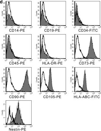
In Stem Cell Res Ther on 4 December 2014 by Wang, Y., Wu, H., et al.
Fig.2.D
-
FC/FACS
-
Homo sapiens (Human)
Collected and cropped from Stem Cell Res Ther by CiteAb, provided under a CC-BY license
Image 1 of 1