Individuals with type 1 diabetes are at increased cardiovascular risk, particularly in the presence of insulin resistance. A prothrombotic environment is believed to contribute to this risk but thrombotic pathways in type 1 diabetes are only partially understood and the role of platelets is incompletely studied. We hypothesised that platelets from individuals with type 1 diabetes exhibit platelet hyperactivity due to both increased propensity for activation and diminished sensitivity to inhibition, with an amplified maladaptive phenotype in those with insulin resistance.
Blood samples were obtained from individuals with type 1 diabetes enrolled on the 'Double diabEtes and adVErse cLinical Outcome: identification of mechanistic Pathways' (DEVELOP) study with insulin resistance assessed as estimated glucose disposal rate (eGDR), whereby eGDR >8 or <6 mg kg-1 min-1 indicates normal insulin sensitivity or advanced insulin resistance, respectively. Platelet function was analysed using whole blood multiparameter flow cytometry to simultaneously measure three distinct markers of activation, including integrin αIIbβ3 (PAC-1 binding), P-selectin (CD62P) and phosphatidylserine (PS) (Annexin V). Both activation and inhibition responses of the platelets were investigated, which were subjected to the machine learning tool Full Annotation Shape-constrained Trees (FAUST) to characterise platelet subpopulations.
A total of 32 individuals with type 1 diabetes were studied (median age [range] of 24 [18-34] years, 59% male, diabetes duration [mean ± SD] of 14.0 ± 6.3 years and HbA1c of 65.3 ± 14.0 mmol/mol [8.1%]). An increased basal expression, measured as mean fluorescence intensity, of all three platelet activation markers was detected in the type 1 diabetes group compared with healthy control participants (CD62P expression 521 ± 246 vs 335 ± 67; p<0.001, PAC-1 370 ± 165 vs 231 ± 88; p=0.011 and PS 869 ± 762 vs 294 ± 109; p=0.001). Following platelet stimulation, an enhanced activation of these markers was found in the type 1 diabetes group. Within the type 1 diabetes group, those with advanced insulin resistance (eGDR<6 mg kg-1 min-1) showed increased platelet activation compared with individuals with normal insulin sensitivity (eGDR>8 mg kg-1 min-1) with single agonist stimulation CD62P expression (29,167 ± 2177 vs 22,829 ± 2535, p<0.001 and PAC-1 19,339 ± 11,749 and 5187 ± 2872, p=0.02). Moreover, individuals with type 1 diabetes showed reduced sensitivity to platelet inhibition by prostacyclin (PGI2) compared with control participants. Stratification of individuals with type 1 diabetes by insulin resistance demonstrated that in the presence of PGI2, suppression of stimulated CD62P was 17 ± 11% and 33 ± 12% (p=0.02) for advanced insulin resistance and normal insulin sensitivity groups, respectively, with even larger differences demonstrated for PAC-1 (48 ± 17% and 75 ± 7%; p=0.006) and PS exposure (33 ± 12% and 84 ± 10%; p=0.001). Furthermore, FAUST analysis showed that, under basal conditions, there was a different distribution of the eight platelet subpopulations comparing advanced insulin resistance and normal insulin sensitivity groups, with differences also detected following PGI2 inhibition.
Our novel characterisation of platelets in type 1 diabetes shows a maladaptive phenotype with increased basal activity together with hyperactivation following stimulation and diminished responses to inhibition. Insulin resistance appears to further drive this adverse thrombotic phenotype, suggesting an enhanced platelet-driven cardiovascular risk in those with type 1 diabetes and reduced insulin sensitivity.
© 2025. The Author(s).
Product Citations: 50
Insulin resistance in type 1 diabetes is a key modulator of platelet hyperreactivity.
In Diabetologia on 30 April 2025 by Sagar, R. C., Yates, D. M., et al.
-
Endocrinology and Physiology
Preprint on BioRxiv : the Preprint Server for Biology on 26 September 2024 by Tranter, J. D., Mikami, R. T., et al.
Platelet shape and volume changes are early mechanical events contributing to platelet activation and thrombosis. Here, we identify single-nucleotide polymorphisms in Leucine-Rich Repeat Containing 8 (LRRC8) protein subunits that form the Volume-Regulated Anion Channel (VRAC) which are independently associated with altered mean platelet volume. LRRC8A is required for functional VRAC in megakaryocytes (MKs) and regulates platelet volume, adhesion, and agonist-stimulated activation, aggregation, ATP secretion and calcium mobilization. MK-specific LRRC8A cKO mice have reduced arteriolar thrombus formation and prolonged arterial thrombosis without affecting bleeding times. Mechanistically, platelet LRRC8A mediates swell-induced ATP/ADP release to amplify agonist-stimulated calcium and PI3K-AKT signaling via P2X1, P2Y 1 and P2Y 12 receptors. Small-molecule LRRC8 channel inhibitors recapitulate defects observed in LRRC8A-null platelets in vitro and in vivo . These studies identify the mechanoresponsive LRRC8 channel complex as an ATP/ADP release channel in platelets which regulates platelet function and thrombosis, providing a proof-of-concept for a novel anti-thrombotic drug target.
-
Genetics
Evaluation of a method to fluorescently label platelets for in-human recovery and survival studies.
In Vox Sanguinis on 1 July 2024 by Feldman, T. P. & Brown, B. L.
Platelets for transfusion are evaluated for in vivo quality using recovery and survival measurements in healthy human subjects. Radiolabelling is the standard for tracing platelets post-transfusion but imposes logistical and technical limitations. This study investigates the in vitro feasibility of labelling platelets with the calcein family of fluorescent dyes as an alternative to radioisotopes or biotin.
Protocols for radiolabelling were adapted for use with calcein acetoxymethyl ester (CAM) and biotin. Labelled platelets were analysed by flow cytometry and evaluated for activation and function. We tested feasibility for labelling without manipulation of platelets and for multiplexing of samples.
Labelling at 2 μg CAM/1010 platelets resulted in >99% of CAM+ platelets. There was no significant difference in activation or aggregation between CAM-labelled or biotinylated platelets and vehicle controls although %CD62P+ was significantly lower in platelets that were not processed for labelling. Addition of CAM to the platelet storage bag labelled >95% of platelets. Platelet populations labelled with different dyes could be distinguished by flow cytometry.
These data provide a rationale for further development of CAM and other fluorescent dyes as tools for measuring post-transfusion kinetics of platelets.
© 2024 International Society of Blood Transfusion.
-
Homo sapiens (Human)
In Frontiers in Immunology on 26 January 2024 by Reusswig, F., Dille, M., et al.
Platelets play an important role in cardiovascular diseases. After acute myocardial infarction, platelets display enhanced activation and migrate into the infarct zone. Furthermore, platelets trigger acute inflammation and cardiac remodeling leading to alterations in scar formation and cardiac function as observed in thrombocytopenic mice. GPVI is the major collagen receptor in platelets and important for platelet activation and thrombus formation and stability. Antibody induced deletion of GPVI at the platelet surface or treatment of mice with recombinant GPVI-Fc results in reduced inflammation and decreased infarct size in a mouse model of AMI. However, the role of GPVI has not been fully clarified to date.
In this study, we found that GPVI is not involved in the inflammatory response in experimental AMI using GPVI deficient mice that were analyzed in a closed-chest model. However, reduced platelet activation in response to GPVI and PAR4 receptor stimulation resulted in reduced pro-coagulant activity leading to improved cardiac remodeling. In detail, GPVI deficiency in mice led to reduced TGF-β plasma levels and decreased expression of genes involved in cardiac remodeling such as Col1a1, Col3a1, periostin and Cthrc1 7 days post AMI. Consequently, collagen quality of the scar shifted to more tight and less fine collagen leading to improved scar formation and cardiac function in GPVI deficient mice at 21d post AMI.
Taken together, this study identifies GPVI as a major regulator of platelet-induced cardiac remodeling and supports the potential relevance of GPVI as therapeutic target to reduce ischemia reperfusion injury and to improve cardiac healing.
Copyright © 2024 Reusswig, Dille, Krüger, Ortscheid, Feige, Gorressen, Fischer and Elvers.
-
Cardiovascular biology
-
Immunology and Microbiology
A randomized clinical trial of bermekimab treatment for clinical improvement of systemic sclerosis.
In IScience on 15 September 2023 by Solomonidi, N., Vlachoyiannopoulos, P. G., et al.
Increased concentrations of interleukin (IL)-1α have been recently described in tissues of patients with systemic sclerosis (SSc) suggesting that IL-1α inhibition may be a target for treatment. We conducted a double-blind, placebo-controlled study to assess the safety and efficacy of the fully humanized IL-1α blocking monoclonal antibody bermekimab in SSc. To evaluate response to treatment, we developed the score of inhibition of progression of SSc which was validated using the CRISS index and the modified CRISS index. The primary endpoint was met in 80% of bermekimab-treated patients vs. 20% of placebo-treated patients (p: 0.023). Most of efficacy was found for increase of carbon monoxide lung diffusion capacity. Production of IL-1α and TNF by circulating mononuclear cells was decreased and the absolute count of CD42/Cd62-platelets was decreased. Results suggest that bermekimab is a promising treatment for SSc.
© 2023 The Authors.
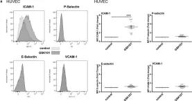
In Physiol Rep on 1 February 2021 by Beddek, K., Raffin, F., et al.
Fig.5.A

-
FC/FACS
-
Homo sapiens (Human)
Collected and cropped from Physiol Rep by CiteAb, provided under a CC-BY license
Image 1 of 3
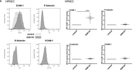
In Physiol Rep on 1 February 2021 by Beddek, K., Raffin, F., et al.
Fig.5.B

-
FC/FACS
-
Homo sapiens (Human)
Collected and cropped from Physiol Rep by CiteAb, provided under a CC-BY license
Image 1 of 3
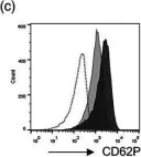
In Br J Pharmacol on 1 September 2020 by Millington-Burgess, S. L., Bonna, A. M., et al.
Fig.2.C

-
FC/FACS
-
Homo sapiens (Human)
Collected and cropped from Br J Pharmacol by CiteAb, provided under a CC-BY license
Image 1 of 3