We previously developed several successful decellularization strategies that yielded porcine cardiac extracellular matrices (pcECMs) exhibiting tissue-specific bioactivity and bioinductive capacity when cultured with various pluripotent and multipotent stem cells. Here, we study the tissue-specific effects of the pcECM on seeded human mesenchymal stem cell (hMSC) phenotypes using reverse transcribed quantitative polymerase chain reaction (RT-qPCR) arrays for cardiovascular related gene expression. We further corroborated interesting findings at the protein level (flow cytometry and immunological stains) as well as bioinformatically using several mRNA sequencing and protein databases of normal and pathologic adult and embryonic (organogenesis stage) tissue expression. We discovered that upon the seeding of hMSCs on the pcECM, they displayed a partial mesenchymal-to-epithelial transition (MET) toward endothelial phenotypes (CD31+) and morphologies, which were preceded by an early spike (~Day 3 onward after seeding) in HAND2 expression at both the mRNA and protein levels compared to that in plate controls. The CRISPR-Cas9 knockout (KO) of HAND2 and its associated antisense long non-coding RNA (HAND2-AS1) regulatory region resulted in proliferation arrest, hypertrophy, and senescent-like morphology. Bioinformatic analyses revealed that HAND2 and HAND2-AS1 are highly correlated in expression and are expressed in many different tissue types albeit at distinct yet tightly regulated expression levels. Deviation (downregulation or upregulation) from these basal tissue expression levels is associated with a long list of pathologies. We thus suggest that HAND2 expression levels may possibly fine-tune hMSCs' plasticity through affecting senescence and mesenchymal-to-epithelial transition states, through yet unknown mechanisms. Targeting this pathway may open up a promising new therapeutic approach for a wide range of diseases, including cancer, degenerative disorders, and aging. Nevertheless, further investigation is required to validate these findings and better understand the molecular players involved, potential inducers and inhibitors of this pathway, and eventually potential therapeutic applications.
Product Citations: 25
In International Journal of Molecular Sciences on 20 November 2023 by Vazana-Netzarim, R., Elmalem, Y., et al.
-
Stem Cells and Developmental Biology
Isolation and characterization of farm pig adipose tissue-derived mesenchymal stromal/stem cells.
In Brazilian Journal of Medical and Biological Research = Revista Brasileira De Pesquisas Médicas E Biológicas / Sociedade Brasileira De Biofísica ... [et Al.] on 9 December 2022 by Garcia, G. A., Oliveira, R. G., et al.
Adipose tissue-derived mesenchymal stromal/stem cells (ASCs) are considered important tools in regenerative medicine and are being tested in several clinical studies. Porcine models are frequently used to obtain adipose tissue, due to the abundance of material and because they have immunological and physiological similarities with humans. However, it is essential to understand the effects and safe application of ASCs from pigs (pASCs) as an alternative therapy for diseases. Although minipigs are easy-to-handle animals that require less food and space, acquiring and maintaining them in a bioterium can be costly. Thus, we present a protocol for the isolation and proliferation of ASCs isolated from adipose tissue of farm pigs. Adipose tissue samples were extracted from the abdominal region of the animals. Because the pigs were not raised in a controlled environment, such as a bioterium, it was necessary to carry out rigorous procedures for disinfection. After this procedure, cells were isolated by mechanical dissociation and enzymatic digestion. A proliferation curve was performed and used to calculate the doubling time of the population. The characterization of pASCs was performed by immunophenotyping and cell differentiation in osteogenic and adipogenic lineages. The described method was efficient for the isolation and cultivation of pASCs, maintaining cellular attributes, such as surface antigens and multipotential differentiation during in vitro proliferation. This protocol presents the isolation and cultivation of ASCs from farm pig as an alternative for the isolation and cultivation of ASCs from minipigs, which require strictly controlled maintenance conditions and a more expensive process.
-
FC/FACS
-
Stem Cells and Developmental Biology
-
Veterinary Research
In Cancers on 18 July 2022 by Kim, D., Kim, J. S., et al.
Cancer-associated fibroblasts (CAFs) reside within the tumor microenvironment, facilitating cancer progression and metastasis via direct and indirect interactions with cancer cells and other stromal cell types. CAFs are composed of heterogeneous subpopulations of activated fibroblasts, including myofibroblastic, inflammatory, and immunosuppressive CAFs. In this study, we sought to identify subpopulations of CAFs isolated from human lung adenocarcinomas and describe their transcriptomic and functional characteristics through single-cell RNA sequencing (scRNA-seq) and subsequent bioinformatics analyses. Cell trajectory analysis of combined total and THY1 + CAFs revealed two branching points with five distinct branches. Based on Gene Ontology analysis, we denoted Branch 1 as "immunosuppressive", Branch 2 as "neoantigen presenting", Branch 4 as "myofibroblastic", and Branch 5 as "proliferative" CAFs. We selected representative branch-specific markers and measured their expression levels in total and THY1 + CAFs. We also investigated the effects of these markers on CAF activity under coculture with lung cancer cells. This study describes novel subpopulations of CAFs in lung adenocarcinoma, highlighting their potential value as therapeutic targets.
-
Cancer Research
In International Journal of Oncology on 1 January 2022 by Watanabe, K., Shiga, K., et al.
The cancer‑stromal interaction has been demonstrated to promote tumor progression, and cancer-associated fibroblasts (CAFs), which are the main components of stromal cells, have attracted attention as novel treatment targets. Chitinase 3-like 1 (CHI3L1) is a chitinase-like protein, which affects cell proliferation and angiogenesis. However, the mechanisms through which cells secrete CHI3L1 and through which CHI3L1 mediates tumor progression in the cancer microenvironment are still unclear. Accordingly, the present study assessed the secretion of CHI3L1 in the microenvironment of colorectal cancer and evaluated how CHI3L1 affects tumor angiogenesis. CAFs and normal fibroblasts (NFs) established from colorectal cancer tissue, and human colon cancer cell lines were evaluated using immunostaining, cytokine antibody array, RNA interference, reverse transcription-quantitative PCR (RT-qPCR), ELISA, western blotting and angiogenesis assays. The expression and secretion of CHI3L1 in CAFs were stronger than those in NFs and colorectal cancer cell lines. In addition, interleukin-13 receptor α2 (IL-13Rα2), a receptor for CHI3L1, was not expressed in colorectal cancer cell lines, but was expressed in fibroblasts, particularly CAFs. Furthermore, the expression and secretion of IL-8 in CAFs was stronger than that in NFs and cancer cell lines, and recombinant CHI3L1 addition increased IL-8 expression in CAFs, whereas knockdown of CHI3L1 suppressed IL-8 expression. Furthermore, IL-13Rα2 knockdown suppressed the enhancement of IL-8 expression induced by CHI3L1 treatment in CAFs. For vascular endothelial growth factor-A (VEGFA), similar results to IL-8 were observed in an ELISA for comparison of secretion between CAFs and NFs and for changes in secretion after CHI3L1 treatment in CAFs; however, no significant differences were observed for changes in expression after CHI3L1 treatment or IL-13Rα2 knockdown in CAFs assessed using RT-qPCR assays. Angiogenesis assays revealed that tube formation in vascular endothelial cells was suppressed by conditioned medium from CAFs with the addition of human CHI3L1 neutralizing antibodies compared with control IgG, and also suppressed by conditioned medium from CAFs transfected with CHI3L1, IL-8 or VEGFA small interfering RNA compared with negative control small interfering RNA. Overall, the present findings indicated that CHI3L1 secreted from CAFs acted on CAFs to increase the secretion of IL-8, thereby affecting tumor angiogenesis in colorectal cancer.
-
ICC-IF
-
Cancer Research
Suppression of Ovarian Cancer Cell Growth by AT-MSC Microvesicles.
In International Journal of Molecular Sciences on 30 November 2020 by Szyposzynska, A., Bielawska-Pohl, A., et al.
Transport of bioactive cargo of microvesicles (MVs) into target cells can affect their fate and behavior and change their microenvironment. We assessed the effect of MVs derived from human immortalized mesenchymal stem cells of adipose tissue-origin (HATMSC2-MVs) on the biological activity of the ovarian cancer cell lines ES-2 (clear cell carcinoma) and OAW-42 (cystadenocarcinoma). The HATMSC2-MVs were characterized using dynamic light scattering (DLS), transmission electron microscopy, and flow cytometry. The anti-tumor properties of HATMSC2-MVs were assessed using MTT for metabolic activity and flow cytometry for cell survival, cell cycle progression, and phenotype. The secretion profile of ovarian cancer cells was evaluated with a protein antibody array. Both cell lines internalized HATMSC2-MVs, which was associated with a decreased metabolic activity of cancer cells. HATMSC2-MVs exerted a pro-apoptotic and/or necrotic effect on ES-2 and OAW-42 cells and increased the expression of anti-tumor factors in both cell lines compared to control. In conclusion, we confirmed an effective transfer of HATMSC2-MVs into ovarian cancer cells that resulted in the inhibition of cell proliferation via different pathways, apoptosis and/or necrosis, which, with high likelihood, is related to the presence of different anti-tumor factors secreted by the ES-2 and OAW-42 cells.
-
Cancer Research
In Cells on 17 March 2020 by Kruminis-Kaszkiel, E., Osowski, A., et al.
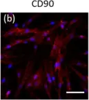
Fig.4.B

-
ICC-IF
-
Homo sapiens (Human)
Collected and cropped from Cells by CiteAb, provided under a CC-BY license
Image 1 of 1
