Fibrosis is defined as an excessive accumulation of extracellular matrix (ECM) components. Many organs are subjected to fibrosis including the lung, liver, heart, skin, kidney, and muscle. Muscle fibrosis occurs in response to trauma, aging, or dystrophies and impairs muscle function. Fibrosis represents a hurdle for the treatment of human muscular dystrophies. While data on the mechanisms of fibrosis have mostly been investigated in mice, dystrophic mouse models often do not recapitulate fibrosis as observed in human patients. Consequently, the cellular and molecular mechanisms that lead to fibrosis in human muscle still need to be identified.
Combining mass cytometry, transcriptome profiling, in vitro co-culture experiments, and in vivo transplantation in immunodeficient mice, we investigated the role and nature of nonmyogenic cells (fibroadipogenic progenitors, FAPs) from human fibrotic muscles of healthy individuals (FibMCT ) and individuals with oculopharyngeal muscular dystrophy (OPMD; FibMOP ), as compared with nonmyogenic cells from human nonfibrotic muscle (MCT ).
We found that the proliferation rate of FAPs from fibrotic muscle is 3-4 times higher than those of FAPs from nonfibrotic muscle (population doubling per day: MCT 0.2 ± 0.1, FibMCT 0.7 ± 0.1, and FibMOP 0.8 ± 0.3). When cocultured with muscle cells, FAPs from fibrotic muscle impair the fusion index unlike MCT FAPs (myoblasts alone 57.3 ± 11.1%, coculture with MCT 43.1 ± 8.9%, with FibMCT 31.7 ± 8.2%, and with FibMOP 36.06 ± 10.29%). We also observed an increased proliferation of FAPs from fibrotic muscles in these co-cultures in differentiation conditions (FibMCT +17.4%, P < 0.01 and FibMOP +15.1%, P < 0.01). This effect is likely linked to the increased activation of the canonical TGFβ-SMAD pathway in FAPs from fibrotic muscles evidenced by pSMAD3 immunostaining (P < 0.05). In addition to the profibrogenic TGFβ pathway, we identified endothelin as a new actor implicated in the altered cross-talk between muscle cells and fibrotic FAPs, confirmed by an improvement of the fusion index in the presence of bosentan, an endothelin receptor antagonist (from 33.8 ± 10.9% to 52.9 ± 10.1%, P < 0.05).
Our data demonstrate the key role of FAPs and their cross-talk with muscle cells through a paracrine signalling pathway in fibrosis of human skeletal muscle and identify endothelin as a new druggable target to counteract human muscle fibrosis.
© 2022 The Authors. Journal of Cachexia, Sarcopenia and Muscle published by John Wiley & Sons Ltd on behalf of Society on Sarcopenia, Cachexia and Wasting Disorders.
Product Citations: 40
In Journal of Cachexia, Sarcopenia and Muscle on 1 June 2022 by Bensalah, M., Muraine, L., et al.
In STAR Protocols on 18 June 2021 by Nishihara, H., Gastfriend, B. D., et al.
We describe the extended endothelial cell culture method (EECM) for the differentiation of human pluripotent stem cells (hPSCs) into brain microvascular endothelial cell (BMEC)-like cells. EECM-BMEC-like cells resemble primary human BMECs in morphology, molecular junctional architecture, and diffusion barrier characteristics. A mature immune phenotype with proper endothelial adhesion molecule expression makes this model distinct from any other hPSC-derived in vitro blood-brain barrier (BBB) model and suitable to study immune cell migration across the BBB in a disease relevant and personalized fashion. For complete details on the use and execution of this protocol, please refer to Lian et al. (2014), Nishihara et al. (2020a).
© 2021 The Author(s).
-
Homo sapiens (Human)
-
Immunology and Microbiology
-
Stem Cells and Developmental Biology
Leukocyte Integrin Antagonists as a Novel Option to Treat Dry Age-Related Macular Degeneration.
In Frontiers in Pharmacology on 16 February 2021 by Baiula, M., Caligiana, A., et al.
Age-related macular degeneration (AMD) is a complex multifactorial degenerative disease that leads to irreversible blindness. AMD affects the macula, the central part of the retina responsible for sharp central vision. Retinal pigment epithelium (RPE) is the main cellular type affected in dry AMD. RPE cells form a monolayer between the choroid and the neuroretina and are in close functional relationship with photoreceptors; moreover, RPE cells are part of the blood retina barrier that is disrupted in ocular diseases such as AMD. During ocular inflammation lymphocytes and macrophages are recruited, contact RPE and produce pro-inflammatory cytokines, which play an important role in AMD pathogenesis. The interaction between RPE and immune cells is mediated by leukocyte integrins, heterodimeric transmembrane receptors, and adhesion molecules, including VCAM-1 and ICAM-1. Within this frame, this study aimed to characterize RPE-leukocytes interaction and to investigate any potentially beneficial effects induced by integrin antagonists (DS-70, MN27 and SR714), developed in previous studies. ARPE-19 cells were co-cultured for different incubation times with Jurkat cells and apoptosis and necrosis levels were analyzed by flow cytometry. Moreover, we measured the mRNA levels of the pro-inflammatory cytokine IL-1β and the expression of adhesion molecules VCAM-1 and ICAM-1. We found that RPE-lymphocyte interaction increased apoptosis and necrosis levels in RPE cells and the expression of IL-1β. This interaction was mediated by the binding of α4β1 and αLβ2 integrins to VCAM-1 and ICAM-1, respectively. The blockade of RPE-lymphocyte interaction with blocking antibodies highlighted the pivotal role played by integrins. Therefore, α4β1 and αLβ2 integrin antagonists were employed to disrupt RPE-lymphocyte crosstalk. Small molecule integrin antagonists proved to be effective in reducing RPE cell death and expression of IL-1β, demonstrating that integrin antagonists could protect RPE cells from detrimental effects induced by the interaction with immune cells recruited to the retina. Overall, the leukocyte integrin antagonists employed in the present study may represent a novel opportunity to develop new drugs to fight dry AMD.
Copyright © 2021 Baiula, Caligiana, Bedini, Zhao, Santino, Cirillo, Gentilucci, Giacomini and Spampinato.
-
Pharmacology
Characterization of zika virus infection of human fetal cardiac mesenchymal stromal cells.
In PLoS ONE on 18 September 2020 by Rossi, F., Josey, B., et al.
Zika virus (ZIKV) is a single-stranded RNA virus belonging to the family Flaviviridae. ZIKV predominantly enters cells using the TAM-family protein tyrosine kinase receptor AXL, which is expressed on a range of cell types, including neural progenitor cells, keratinocytes, dendritic cells, and osteoblasts. ZIKV infections have been associated with fetal brain damage, which prompted the World Health Organization to declare a public health emergency in 2016. ZIKV infection has also been linked to birth defects in other organs. Several studies have reported congenital heart defects (CHD) in ZIKV infected infants and cardiovascular complications in adults infected with ZIKV. To develop a better understanding of potential causes for these pathologies at a cellular level, we characterized ZIKV infection of human fetal cardiac mesenchymal stromal cells (fcMSCs), a cell type that is known to contribute to both embryological development as well as adult cardiac physiology. Total RNA, supernatants, and/or cells were collected at various time points post-infection to evaluate ZIKV replication, cell death, and antiviral responses. We found that ZIKV productively infected fcMSCs with peak (~70%) viral mRNA detected at 48 h. Use of an antibody blocking the AXL receptor decreased ZIKV infection (by ~50%), indicating that the receptor is responsible to a large extent for viral entry into the cell. ZIKV also altered protein expression of several mesenchymal cell markers, which suggests that ZIKV could affect fcMSCs' differentiation process. Gene expression analysis of fcMSCs exposed to ZIKV at 6, 12, and 24 h post-infection revealed up-regulation of genes/pathways associated with interferon-stimulated antiviral responses. Stimulation of TLR3 (using poly I:C) or TLR7 (using Imiquimod) prior to ZIKV infection suppressed viral replication in a dose-dependent manner. Overall, fcMSCs can be a target for ZIKV infection, potentially resulting in CHD during embryological development and/or cardiovascular issues in ZIKV infected adults.
-
FC/FACS
-
Cardiovascular biology
-
Immunology and Microbiology
In International Journal of Molecular Sciences on 21 March 2020 by Kim, J. H., Lim, I. R., et al.
Thymosin β4 (Tβ4) is a G-actin sequestering protein that contributes to diverse cellular activities, such as migration and angiogenesis. In this study, the beneficial effects of combined cell therapy with Tβ4 and human adipose-derived stem cells (hASCs) in a mouse ischemic hindlimb model were investigated. We observed that exogenous treatment with Tβ4 enhanced endogenous TMSB4X mRNA expression and promoted morphological changes (increased cell length) in hASCs. Interestingly, Tβ4 induced the active state of hASCs by up-regulating intracellular signaling pathways including the PI3K/AKT/mTOR and MAPK/ERK pathways. Treatment with Tβ4 significantly increased cell migration and sprouting from microbeads. Moreover, additional treatment with Tβ4 promoted the endothelial differentiation potential of hASCs by up-regulating various angiogenic genes. To evaluate the in vivo effects of the Tβ4-hASCs combination on vessel recruitment, dorsal window chambers were transplanted, and the co-treated mice were found to have a significantly increased number of microvessel branches. Transplantation of hASCs in combination with Tβ4 was found to improve blood flow and attenuate limb or foot loss post-ischemia compared to transplantation with hASCs alone. Taken together, the therapeutic application of hASCs combined with Tβ4 could be effective in enhancing endothelial differentiation and vascularization for treating hindlimb ischemia.
-
IF
-
Stem Cells and Developmental Biology
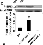
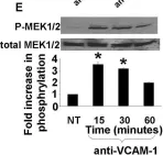
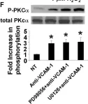
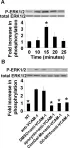
In PLoS One on 28 October 2011 by Abdala-Valencia, H., Berdnikovs, S., et al.
Fig.4.C

-
WB
-
Collected and cropped from PLoS One by CiteAb, provided under a CC-BY license
Image 1 of 4
In PLoS One on 28 October 2011 by Abdala-Valencia, H., Berdnikovs, S., et al.
Fig.4.E

-
WB
-
Collected and cropped from PLoS One by CiteAb, provided under a CC-BY license
Image 1 of 4
In PLoS One on 28 October 2011 by Abdala-Valencia, H., Berdnikovs, S., et al.
Fig.4.F

-
WB
-
Collected and cropped from PLoS One by CiteAb, provided under a CC-BY license
Image 1 of 4
In PLoS One on 28 October 2011 by Abdala-Valencia, H., Berdnikovs, S., et al.
Fig.5.A

-
WB
-
Collected and cropped from PLoS One by CiteAb, provided under a CC-BY license
Image 1 of 4