In celiac disease (CeD), interleukin 15 (IL-15) affects the epithelial barrier by acting on intraepithelial lymphocytes, promoting interferon γ (IFN-γ) production and inducing strong cytotoxic activity as well as eliciting apoptotic death of enterocytes by the Fas/Fas ligand system. This study investigates the effects of a monoclonal antibody neutralizing the effects of IL-15 (aIL-15) on tissue-damaging immune response in untreated CeD patients by using an organ culture system. Jejunal biopsies from 10 untreated CeD patients were cultured ex vivo with or without aIL-15. Epithelial expressions of CD95/Fas, HLA-E and perforin were analyzed by immunohistochemistry. Apoptosis was detected in the epithelium by using the terminal deoxynucleotidyl transferase-mediated dUTP nick-end labeling (TUNEL) assay. Additionally, the surface epithelium compartment of ex vivo cultured biopsy samples was isolated by laser capture microdissection (LCM). RNA from each LCM sample was extracted and the relative expression of IFN-γ was evaluated by quantitative reverse transcriptase-PCR (qRT-PCR). Biopsies cultured with the aIL-15 antibody showed a reduction in Fas, HLA-E and perforin epithelial expression, as well as a decrease in epithelial TUNEL+ cells compared to biopsies cultured without the aIL-15 antibody. Moreover, downregulation of epithelial IFN-γ expression was recorded in biopsies incubated with aIL-15, compared to those cultured without aIL-15. Our findings suggest that neutralizing the effects of IL-15 in ex vivo cultured untreated CeD intestinal mucosa could block apoptosis by downregulating Fas and HLA-E expression and the release of cytotoxic proteins, such as perforin. Furthermore, it can dampen the hyperactive immune response by reducing IFN-γ expression. More generally, our study provides new evidence for the effects of anti-IL-15 neutralizing monoclonal antibodies in preventing or repairing epithelial damage and further supports the concept that IL-15 is a meaningful therapeutic target in CeD, or inflammatory diseases associated with the upregulation of IL-15.
Product Citations: 61
In Cells on 6 February 2025 by Rotondi Aufiero, V., Iacomino, G., et al.
-
IHC
-
Homo sapiens (Human)
-
Cell Biology
-
Immunology and Microbiology
In Frontiers in Immunology on 6 July 2021 by Huot, N., Rascle, P., et al.
CD4 T cell responses constitute an important component of adaptive immunity and are critical regulators of anti-microbial protection. CD4+ T cells expressing CD32a have been identified as a target for HIV. CD32a is an Fcγ receptor known to be expressed on myeloid cells, granulocytes, B cells and NK cells. Little is known about the biology of CD32+CD4+ T cells. Our goal was to understand the dynamics of CD32+CD4+ T cells in tissues. We analyzed these cells in the blood, lymph nodes, spleen, ileum, jejunum and liver of two nonhuman primate models frequently used in biomedical research: African green monkeys (AGM) and macaques. We studied them in healthy animals and during viral (SIV) infection. We performed phenotypic and transcriptomic analysis at different stages of infection. In addition, we compared CD32+CD4+ T cells in tissues with well-controlled (spleen) and not efficiently controlled (jejunum) SIV replication in AGM. The CD32+CD4+ T cells more frequently expressed markers associated with T cell activation and HIV infection (CCR5, PD-1, CXCR5, CXCR3) and had higher levels of actively transcribed SIV RNA than CD32-CD4+T cells. Furthermore, CD32+CD4+ T cells from lymphoid tissues strongly expressed B-cell-related transcriptomic signatures, and displayed B cell markers at the cell surface, including immunoglobulins CD32+CD4+ T cells were rare in healthy animals and blood but increased strongly in tissues with ongoing viral replication. CD32+CD4+ T cell levels in tissues correlated with viremia. Our results suggest that the tissue environment induced by SIV replication drives the accumulation of these unusual cells with enhanced susceptibility to viral infection.
Copyright © 2021 Huot, Rascle, Planchais, Contreras, Passaes, Le Grand, Beignon, Kornobis, Legendre, Varet, Saez-Cirion, Mouquet, Jacquelin and Müller-Trutwin.
-
Immunology and Microbiology
In International Journal of Molecular Sciences on 27 August 2020 by Dhupar, R., Okusanya, O. T., et al.
While T cell-based cancer immunotherapies have shown great promise, there remains a need to understand how individual metastatic tumor environments impart local T cell dysfunction. At advanced stages, cancers that metastasize to the pleural space can result in a malignant pleural effusion (MPE) that harbors abundant tumor and immune cells, often exceeding 108 leukocytes per liter. Unlike other metastatic sites, MPEs are readily and repeatedly accessible via indwelling catheters, providing an opportunity to study the interface between tumor dynamics and immunity. In the current study, we examined CD8+ T cells within MPEs collected from patients with heterogeneous primary tumors and at various stages in treatment to determine (1) if these cells possess anti-tumor activity following removal from the MPE, (2) factors in the MPE that may contribute to their dysfunction, and (3) the phenotypic changes in T cell populations that occur following ex vivo expansion. Co-cultures of CD8+ T cells with autologous CD45- tumor containing cells demonstrated cytotoxicity (p = 0.030) and IFNγ production (p = 0.003) that inversely correlated with percent of myeloid derived suppressor cells, lactate, and lactate dehydrogenase (LDH) within the MPE. Ex vivo expansion of CD8+ T cells resulted in progressive differentiation marked by distinct populations expressing decreased CD45RA, CCR7, CD127, and increased inhibitory receptors. These findings suggest that MPEs may be a source of tumor-reactive T cells and that the cellular and acellular components suppress optimal function.
-
Cancer Research
-
Immunology and Microbiology
B cells and tertiary lymphoid structures promote immunotherapy response.
In Nature on 1 January 2020 by Helmink, B. A., Reddy, S. M., et al.
Treatment with immune checkpoint blockade (ICB) has revolutionized cancer therapy. Until now, predictive biomarkers1-10 and strategies to augment clinical response have largely focused on the T cell compartment. However, other immune subsets may also contribute to anti-tumour immunity11-15, although these have been less well-studied in ICB treatment16. A previously conducted neoadjuvant ICB trial in patients with melanoma showed via targeted expression profiling17 that B cell signatures were enriched in the tumours of patients who respond to treatment versus non-responding patients. To build on this, here we performed bulk RNA sequencing and found that B cell markers were the most differentially expressed genes in the tumours of responders versus non-responders. Our findings were corroborated using a computational method (MCP-counter18) to estimate the immune and stromal composition in this and two other ICB-treated cohorts (patients with melanoma and renal cell carcinoma). Histological evaluation highlighted the localization of B cells within tertiary lymphoid structures. We assessed the potential functional contributions of B cells via bulk and single-cell RNA sequencing, which demonstrate clonal expansion and unique functional states of B cells in responders. Mass cytometry showed that switched memory B cells were enriched in the tumours of responders. Together, these data provide insights into the potential role of B cells and tertiary lymphoid structures in the response to ICB treatment, with implications for the development of biomarkers and therapeutic targets.
-
Immunology and Microbiology
In Frontiers in Immunology on 21 May 2019 by Herranz, G., Aguilera, P., et al.
Multivesicular bodies (MVB) are endocytic compartments that enclose intraluminal vesicles (ILVs) formed by inward budding from the limiting membrane of endosomes. In T lymphocytes, ILVs are secreted as Fas ligand-bearing, pro-apoptotic exosomes following T cell receptor (TCR)-induced fusion of MVB with the plasma membrane at the immune synapse (IS). In this study we show that protein kinase C δ (PKCδ), a novel PKC isotype activated by diacylglycerol (DAG), regulates TCR-controlled MVB polarization toward the IS and exosome secretion. Concomitantly, we demonstrate that PKCδ-interfered T lymphocytes are defective in activation-induced cell death. Using a DAG sensor based on the C1 DAG-binding domain of PKCδ and a GFP-PKCδ chimera, we reveal that T lymphocyte activation enhances DAG levels at the MVB endomembranes which mediates the association of PKCδ to MVB. Spatiotemporal reorganization of F-actin at the IS is inhibited in PKCδ-interfered T lymphocytes. Therefore, we propose PKCδ as a DAG effector that regulates the actin reorganization necessary for MVB traffic and exosome secretion.
-
Cell Biology
-
Immunology and Microbiology
-
Neuroscience
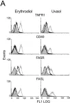
In PLoS One on 23 June 2009 by Martín, R., Ibeas, E., et al.
Fig.7.A

-
FC/FACS
-
Homo sapiens (Human)
Collected and cropped from PLoS One by CiteAb, provided under a CC-BY license
Image 1 of 1