Proteasome inhibitors (PIs) bortezomib, carfilzomib and ixazomib are approved for the treatment of multiple myeloma and mantle cell lymphoma and have clinical activity in acute lymphoblastic leukemia (ALL). The predominant form of proteasome in these hematologic malignancies is the lymphoid tissue-specific immunoproteasome. FDA-approved PIs inhibit immunoproteasomes and ubiquitously expressed constitutive proteasomes causing on-target toxicities in non-hematological tissues. Replacing PIs with selective immunoproteasome inhibitors (IPIs) should reduce these toxicities. We have previously shown that IPI ONX-0914 causes apoptosis of ALL cells expressing the KMT2A::AFF1 (MLL-AF4) fusion protein but did not elucidate the mechanism. Here we show that a novel, highly specific IPI M3258 induces rapid apoptosis in ALL cells in vitro and is comparable to bortezomib in its ability to reduce tumor growth and to cause tumor regression when combined with chemotherapy in vivo. Treatment of KMT2A::AFF1 ALL cells with M3258, ONX-0914, and bortezomib induced proteotoxic stress that was prevented by the protein synthesis inhibitor cycloheximide, which dramatically desensitized cells to PI-induced apoptosis. Thus, similar to multiple myeloma, ALL cells are sensitive to PIs and IPIs due to increased proteotoxic stress caused by elevated rates of protein synthesis.
© 2025. The Author(s).
Product Citations: 224
In Scientific Reports on 19 May 2025 by Jenkins, T. W., Fitzgerald, J. E., et al.
-
Cancer Research
In Scientific Reports on 5 March 2025 by Morris, K., Masri, S., et al.
Platelet-cancer cell interactions play a significant role in metastasis. Indeed, they interact via a plethora of receptors, including integrins (e.g. ⍺IIbβ3 and ⍺vβ3), and calcium is essential for both their stability and function. Additionally, calcium plays a significant role in the coagulation cascade, and the implication of calcium level changes on metastatic dissemination and cancer-associated thrombosis are not fully understood. A significant proportion of cancer patients suffer from hypercalcemia and have a worse prognosis. We hypothesized that calcium levels are important for platelet-cancer cell interactions that are mediated via integrins, thus this can be leveraged to disrupt platelet support to the metastatic process. In this study, we assessed the detection of integrins ⍺IIbβ3 and ⍺vβ3 on platelets and cancer cells, platelet function, and the respective receptors implicated in platelet function, while modulating calcium levels. The effect of calcium levels on platelet-cancer cell interactions and cancer cell invasion in vitro was also assessed. Our data demonstrates that calcium levels affect surface integrins, and receptors involved in platelet-cancer cell interactions. In addition, calcium levels significantly affect platelet activation and aggregation. In our experimental scenarios, calcium depletion modulates platelet-cancer cell interaction with MDA-MB-231 breast cancer cells, while hypercalcemic environments did not affect interaction. Meanwhile, hypercalcemia leads to enhanced cancer cell invasion for both MDA-MB-231 and A549 cells in the presence of platelets. Thus, this study provides a greater understanding of the dynamics associated with the effects of calcium and platelet-cancer cell interactions mediated by integrins.
© 2025. The Author(s).
-
Cancer Research
In Regenerative Therapy on 1 March 2025 by Morita, K., Nakayama, M., et al.
Tissue engineering plays a pivotal role in the advancement of regenerative medicine. Thermoresponsive culture dishes, coated with specialized polymers that control cell adhesion through temperature fluctuations, enable the processing of cells into sheets for medical applications while maintaining their intact state. Cell sheets prepared using these culture dishes have been incorporated into several commercial pharmaceutical products. However, controlling the detachability of cell sheets using conventional thermoresponsive culture dishes remains a challenge, and often leads to unexpected detachment during cultivation. In this study, we developed a thermoresponsive culture dish with tunable cell detachability using a thermoresponsive block copolymer, poly(butyl methacrylate)-b-poly(N-isopropylacrylamide) (PBMA-PIPAAm), which is a specialized polymer that allows precise control of the amount of surface-immobilized polymer and polymer layer thickness. Culturing periodontal ligament-derived mesenchymal stem cells on these dishes demonstrated fully tunable detachability without compromising cell properties compared to conventional thermoresponsive dishes (UpCell®). Thermoresponsive PBMA-PIPAAm-coated culture dishes enable the complete on-demand detachment of transplantable cell sheets, thereby avoiding unexpected detachment that may increase production costs and reduce technical hurdles in the manufacturing process. The PBMA-PIPAAm coating method has the potential to contribute to biomedical and clinical applications of mesenchymal stem cell sheets.
© 2025 The Author(s).
-
Stem Cells and Developmental Biology
In International Journal of Molecular Sciences on 11 February 2025 by David, P., Kouhestani, D., et al.
The poor prognosis of pancreatic ductal adenocarcinoma (PDAC) is largely due to several challenges, such as late diagnosis, early metastasis, limited response to chemotherapy, aggressive tumor biology, and high rates of tumor recurrence. Therefore, the development of a non-invasive and effective method for early detection of PDAC is crucial to improving patient outcomes. Continued research and exploration in this area are essential to enhance early detection methods and ultimately improve the prognosis for individuals with PDAC. In this study, we examined 37 exosomal surface proteins through a multiplex flow cytometry test on peripheral plasma samples from a group of 51 clinical control individuals (including healthy volunteers and non-cancer patients (Cholecystectomy, Hernia, healthy volunteers)), 21 pancreatitis, and 48 patients diagnosed with PDAC. Our research findings revealed that the level of exosomal CD40 expression is significantly lower in patients with PDAC and pancreatitis compared to non-cancer patients (p < 0.0001). Additionally, pancreatitis patients exhibited higher levels of exosomal CD25 expression than PDAC patients (p = 0.0104). PDAC patients with higher exo-CD40 had worse survival than patients with lower exo-CD40 (p = 0.0035). Similarly, PDAC patients with higher exo-CD25 showed worse survival in comparison to patients with lower exo-CD25 (p = 0.04). Statistical analysis revealed that exosomal CD40 achieved an AUC of 0.827 in distinguishing PDAC from clinical controls. Combining exo-CD40 along with exo-CD25 and CA19-9 discriminated PDAC patients from clinical controls with an AUC of 0.92. Exo-CD40 and exo-CD25 proteins found in exosomes isolated from plasma can serve as excellent non-invasive biomarkers for the early diagnosis of PDAC. Further larger scale studies are needed to validate combined exo-CD40 and exo-CD25 as a diagnostic tool for the identification of PDAC patients through non-invasive liquid biopsy.
-
Cancer Research
In Frontiers in Immunology on 7 February 2025 by Tomasello, L., Coppola, A., et al.
The primary outcome was the evaluation of the T-cell phenotype in autoimmune primary adrenal insufficiency (PAI). Secondary outcomes included the evaluation of the CD4+CD25+Foxp3+ Treg population and the gene expression levels of IL-6, IL-17A, cyclooxygenase (COX)-2, heat shock proteins (HSP)-70, indoleamine-2,3-dioxygenase (IDO), programmed death-ligand 1 (PD-L1), inducible nitric oxide synthase (iNOS), and thioredoxin (TXN)-1.
We prospectively included 15 patients with PAI on conventional glucocorticoid (GC) replacement therapy, 15 switched to dual-release hydrocortisone (DR-HC), and 20 healthy controls. Serum inflammatory parameters and peripheral blood mononuclear cells (PBMCs) were evaluated at baseline and after 12 months of treatment.
At baseline, significantly higher CD4+ and CD8+ (both p < 0.001) T-cell percentages, a lower CD4+/CD8+ ratio (p < 0.05), and higher CD25+ and CD4+/CD25+ T cells (both p < 0.001) were observed in PAI compared to controls. After 12 months of DR-HC treatment, we found significantly lower IL-6 (p = 0.019), IL-17A (p = 0.046), COX-2 (p < 0.001), HSP-70 (p = 0.006), and TXN-1 (p = 0.008) and higher PD-L1 (p < 0.001) and IDO (p < 0.001) mRNA values compared to baseline. After 12 months of DR-HC treatment, a significant increase in CD4+ T cells (p = 0.012), PD-L1 (p = 0.003), and IDO (p < 0.001) and a decrease in CD8+ T cells (p < 0.001), IL-6 (p = 0.003), IL-17A (p = 0.0014), COX-2 (p < 0.001), HSP-70 (p = 0.005), and TXN-1 (p = 0.0008), as well as a significantly higher conversion in the CD4+/CD8+ ratio (p = 0.033), were observed compared to conventional GCs.
The switch from conventional GCs to DR-HC treatment altered the T lymphocyte phenotype and CD4+/CD8+ ratio in a Treg-independent manner, inducing significant improvements in the immune and inflammatory profile in PAI.
Copyright © 2025 Tomasello, Coppola, Pizzolanti, Giordano, Arnaldi and Guarnotta.
-
Homo sapiens (Human)
-
Cardiovascular biology
-
Immunology and Microbiology
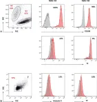
In TH Open on 1 October 2019 by Papageorgiou, L., Alhaj Hussen, K., et al.
Fig.2.A

-
FC/FACS
-
Homo sapiens (Human)
Collected and cropped from TH Open by CiteAb, provided under a CC-BY license
Image 1 of 1