Vacuolar protein sorting 41 (VPS41), a component of the homotypic fusion and protein sorting (HOPS) complex for lysosomal fusion, is essential for the trafficking of lysosomal membrane proteins via lysosome-associated membrane protein (LAMP) carriers from the trans-Golgi network (TGN) to endo/lysosomes. However, the molecular mechanisms underlying this pathway and VPS41's role herein remain poorly understood. Here, we investigated the effects of ectopically localizing VPS41 to mitochondria on LAMP distribution. Using electron microscopy, we identified that mitochondrial-localized VPS41 recruited LAMP1- and LAMP2A-positive vesicles resembling LAMP carriers. The retention using selective hooks (RUSH) system further revealed that newly synthesized LAMPs were specifically recruited by mitochondrial VPS41, a function not shared by other HOPS subunits. Notably, we identified the small GTPase Arl8b as a critical factor for LAMP carrier trafficking. Arl8b was present on LAMP carriers and bound to the WD40 domain of VPS41, enabling their recruitment. These findings reveal a unique role of VPS41 in recruiting TGN-derived LAMP carriers and expand our understanding of VPS41-Arl8b interactions beyond endosome-lysosome fusion, providing new insights into lysosomal trafficking mechanisms.
© 2025 Sanzà et al.
Product Citations: 67
VPS41 recruits biosynthetic LAMP-positive vesicles through interaction with Arl8b.
In The Journal of Cell Biology on 3 April 2025 by Sanzà, P., van der Beek, J., et al.
-
Cell Biology
In Antiviral Research on 1 June 2024 by Glitscher, M., Benz, N. I., et al.
Zoonoses such as ZIKV and SARS-CoV-2 pose a severe risk to global health. There is urgent need for broad antiviral strategies based on host-targets filling gaps between pathogen emergence and availability of therapeutic or preventive strategies. Significant reduction of pathogen titers decreases spread of infections and thereby ensures health systems not being overloaded and public life to continue. Based on previously observed interference with FGFR1/2-signaling dependent impact on interferon stimulated gene (ISG)-expression, we identified Pim kinases as promising druggable cellular target. We therefore focused on analyzing the potential of pan-Pim kinase inhibition to trigger a broad antiviral response. The pan-Pim kinase inhibitor AZD1208 exerted an extraordinarily high antiviral effect against various ZIKV isolates, SARS-CoV-2 and HBV. This was reflected by strong reduction in viral RNA, proteins and released infectious particles. Especially in case of SARS-CoV-2, AZD1208 led to a complete removal of viral traces in cells. Kinome-analysis revealed vast changes in kinase landscape upon AZD1208 treatment, especially for inflammation and the PI3K/Akt-pathway. For ZIKV, a clear correlation between antiviral effect and increase in ISG-expression was observed. Based on a cell culture model with impaired ISG-induction, activation of the PI3K-Akt-mTOR axis, leading to major changes in the endolysosomal equilibrium, was identified as second pillar of the antiviral effect triggered by AZD1208-dependent Pim kinase inhibition, also against HBV. We identified Pim-kinases as cellular target for a broad antiviral activity. The antiviral effect exerted by inhibition of Pim kinases is based on at least two pillars: innate immunity and modulation of the endolysosomal system.
Copyright © 2024 The Authors. Published by Elsevier B.V. All rights reserved.
-
Immunology and Microbiology
In Cellular and Molecular Gastroenterology and Hepatology on 9 January 2024 by Glitscher, M., Spannaus, I. M., et al.
A peculiar feature of the hepatitis E virus (HEV) is its reliance on the exosomal route for viral release. Genomic replication is mediated via the viral polyprotein pORF1, yet little is known about its subcellular localization.
Subcellular localization of pORF1 and its subdomains, generated and cloned based on a structural prediciton of the viral replicase, was analyzed via confocal laser scanning microscopy. Exosomes released from cells were isolated via ultracentrifugation and analyzed by isopycnic density gradient centrifugation. This was followed by fluorimetry or Western blot analyses or reverse transcriptase-polymerase chain reaction to analyze separated particles in more detail.
We found pORF1 to be accumulating within the endosomal system, most dominantly to multivesicular bodies (MVBs). Expression of the polyprotein's 7 subdomains revealed that the papain-like cysteine-protease (PCP) is the only domain localizing like the full-length protein. A PCP-deficient pORF1 mutant lost its association to MVBs. Strikingly, both pORF1 and PCP can be released via exosomes. Similarly, genomic RNA still is released via exosomes in the absence of pORF2/3.
Taken together, we found that pORF1 localizes to MVBs in a PCP-dependent manner, which is followed by exosomal release. This reveals new aspects of HEV life cycle, because replication and release could be coupled at the endosomal interface. In addition, this may mediate capsid-independent spread or may facilitate the spread of viral infection, because genomes entering the cell during de novo infection readily encounter exosomally transferred pORF1.
Copyright © 2024 The Authors. Published by Elsevier Inc. All rights reserved.
In Human Genetics on 1 March 2023 by Smits, D. J., Dekker, J., et al.
CLEC16A is a membrane-associated C-type lectin protein that functions as a E3-ubiquitin ligase. CLEC16A regulates autophagy and mitophagy, and reportedly localizes to late endosomes. GWAS studies have associated CLEC16A SNPs to various auto-immune and neurological disorders, including multiple sclerosis and Parkinson disease. Studies in mouse models imply a role for CLEC16A in neurodegeneration. We identified bi-allelic CLEC16A truncating variants in siblings from unrelated families presenting with a severe neurodevelopmental disorder including microcephaly, brain atrophy, corpus callosum dysgenesis, and growth retardation. To understand the function of CLEC16A in neurodevelopment we used in vitro models and zebrafish embryos. We observed CLEC16A localization to early endosomes in HEK293T cells. Mass spectrometry of human CLEC16A showed interaction with endosomal retromer complex subunits and the endosomal ubiquitin ligase TRIM27. Expression of the human variant leading to C-terminal truncated CLEC16A, abolishes both its endosomal localization and interaction with TRIM27, suggesting a loss-of-function effect. CLEC16A knockdown increased TRIM27 adhesion to early endosomes and abnormal accumulation of endosomal F-actin, a sign of disrupted vesicle sorting. Mutagenesis of clec16a by CRISPR-Cas9 in zebrafish embryos resulted in accumulated acidic/phagolysosome compartments, in neurons and microglia, and dysregulated mitophagy. The autophagocytic phenotype was rescued by wild-type human CLEC16A but not the C-terminal truncated CLEC16A. Our results demonstrate that CLEC16A closely interacts with retromer components and regulates endosomal fate by fine-tuning levels of TRIM27 and polymerized F-actin on the endosome surface. Dysregulation of CLEC16A-mediated endosomal sorting is associated with neurodegeneration, but it also causes accumulation of autophagosomes and unhealthy mitochondria during brain development.
© 2022. The Author(s).
-
Cell Biology
-
Genetics
In Molecular Biology of the Cell on 1 August 2022 by Kennedy, A., Ren, H. Y., et al.
We report on how the endoplasmic reticulum (ER)-associated-autophagy pathway (ERAA) delivers P23H-rhodopsin (P23H-R) to the lysosome. P23H-R accumulates in an ERAD-resistant conformation that is stabilized in a detergent-soluble state by DNAJB12 and Hsp70. P23H-R, DNAJB12, and FIP200 colocalize in discrete foci that punctuate the rim of omegasome rings coated by WIPI1. Loss of DNAJB12 function prevents the association of P23H-R containing ER tubules with omegasomes. P23H-R tubules thread through the wall of WIPI1 rings into their central cavity. Transfer of P23H-R from ER-connected phagophores to lysosomes requires GABARAP and is associated with the transient docking of lysosomes to WIPI1 rings. After departure from WIPI1 rings, new patches of P23H-R are seen in the membranes of lysosomes. The absence of GABARAP prevents transfer of P23H-R from phagophores to lysosomes without interfering with docking. These data identify lysosome docking to omegasomes as an important step in the DNAJB12- and GABARAP-dependent autophagic disposal of dominantly toxic P23H-R.
-
Cell Biology
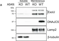
In Nat Commun on 29 June 2021 by Deruelle, V., Bouillot, S., et al.
Fig.3.A

-
WB
-
Collected and cropped from Nat Commun by CiteAb, provided under a CC-BY license
Image 1 of 3
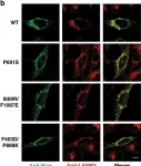
In Nat Commun on 9 January 2020 by Long, T., Qi, X., et al.
Fig.6.B

-
ICC-IF
-
Collected and cropped from Nat Commun by CiteAb, provided under a CC-BY license
Image 1 of 3
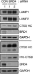
In Mol Cell on 18 May 2017 by Sakamaki, J. I., Wilkinson, S., et al.
Fig.3.B

-
WB
-
Homo sapiens (Human)
Collected and cropped from Mol Cell by CiteAb, provided under a CC-BY license
Image 1 of 3