
Mesenchymal stem cells (MSCs) are frequently used for therapeutic applications in both pre-clinical and clinical settings owing to their capacity for immune modulation and neuroprotective effects. However, transient fever is commonly observed as an adverse event following MSC injection in patients with Alzheimer's disease (AD). In this study, we investigated the potential impact of immunosuppressants such as dexamethasone and tacrolimus on altering the characteristics of human mesenchymal stem cells (hMSCs). Additionally, we examined whether these immunosuppressants affect the persistence of hMSCs or the immune response upon their administration into the brain parenchyma of AD mice. The exposure of hMSCs to high concentrations of dexamethasone and tacrolimus in vitro did not significantly alter the characteristics of hMSCs. The expression of genes related to innate immune responses, such as Irak1, Irf3, Nod1, and Ifnar1, was significantly downregulated by the additional administration of dexamethasone and tacrolimus to the brain parenchyma of AD mice. However, hMSC persistence in the AD mouse brain was not affected. The results of this study support the use of immunosuppressants to mitigate fever during stem cell therapy in patients with AD.
Product Citations: 112
In International Journal of Stem Cells on 30 May 2025 by Lee, N. K., Na, D. L., et al.
-
Stem Cells and Developmental Biology
In Cellular Molecular Immunology on 1 April 2025 by Xu, X., Xu, P., et al.
Macrophage polarization and energy metabolic reprogramming play pivotal roles in the onset and progression of inflammatory arthritis. Moreover, although previous studies have reported that the proviral integration of Moloney virus 2 (Pim2) kinase is involved in various cancers through the mediation of aerobic glycolysis in cancer cells, its role in inflammatory arthritis remains unclear. In this study, we demonstrated that multiple metabolic enzymes are activated upon Pim2 upregulation during M1 macrophage polarization. Specifically, Pim2 directly phosphorylates PGK1-S203, PDHA1-S300, and PFKFB2-S466, thereby promoting glycolytic reprogramming. Pim2 expression was elevated in macrophages from patients with inflammatory arthritis and collagen-induced arthritis (CIA) model mice. Conditional knockout of Pim2 in macrophages or administration of the Pim2 inhibitor HJ-PI01 attenuated arthritis development by inhibiting M1 macrophage polarization. Through molecular docking and dynamic simulation, bexarotene was identified as an inhibitor of Pim2 that inhibits glycolysis and downstream M1 macrophage polarization, thereby mitigating the progression of inflammatory arthritis. For targeted treatment, neutrophil membrane-coated bexarotene (Bex)-loaded PLGA-based nanoparticles (NM@NP-Bex) were developed to slow the progression of inflammatory arthritis by suppressing the polarization of M1 macrophages, and these nanoparticles (NPs) exhibited superior therapeutic effects with fewer side effects. Taken together, the results of our study demonstrated that targeting Pim2 inhibition could effectively alleviate inflammatory arthritis via glycolysis inhibition and reversal of the M1/M2 macrophage imbalance. NM@NPs loaded with bexarotene could represent a promising targeted strategy for the treatment of inflammatory arthritis.
© 2025. The Author(s).
-
Biochemistry and Molecular biology
-
Cell Biology
-
Immunology and Microbiology
In Experimental Hematology Oncology on 17 February 2025 by Caratelli, S., De Paolis, F., et al.
Recent studies have shown that CD32/CD8a/CD28/CD3ζ chimeric receptor cells directly kill breast cancer cells, suggesting the existence of cell surface myeloid FcγR alternative ligands (ALs). Here, we investigated the metabolism, ALs, cytotoxicity, and immunoregulatory functions of CD64/CD28/CD3ζ in colorectal cancer (CRC) and squamous cell carcinoma of the head and neck.
The CD64/CD28/CD3ζ -SFG retroviral vector was used to produce viruses for T-cell transduction. T-cell expansion and differentiation were monitored via flow cytometry. Gene expression was assessed by RNA-seq. Bioenergetics were documented on a Seahorse extracellular flux analyzer. CD64/CD28/CD3ζ polarization was identified via confocal microscopy. Cytotoxicity was determined by MTT assay and bioluminescent imaging, and flow cytometry. Tridimensional antitumor activity of CD64/CD28/CD3ζ T cells was achieved by utilizing HCT116-GFP 3D spheroids via the IncuCyte S3 Live-Cell Analysis system. The intraperitoneal distribution and antitumor activity of NIR-CD64/CD28/CD3ζ and NIR-nontransduced T cells were investigated in CB17-SCID mice bearing subcutaneous FaDu Luc + cells by bioluminescent and fluorescent imaging. IFNγ was assessed by ELISA.
Compared to CD16/CD8a/CD28/CD3ζ T cells, CD32/CD8a/CD28/CD3ζ T cells, and non-transduced T cells, CD64/CD28/CD3ζ T cells exhibited the highest levels of cell expansion and persistence capacity. A total of 235 genes linked to cell division and 52 genes related to glycolysis were overexpressed. The glycolytic phenotype was confirmed by functional in vitro studies accompanied by preferential T-cell effector memory differentiation. Interestingly, oxamic acid was found to inhibit CD64-CR T cell proliferation, indicating the involvement of lactate. Upon CD64/CD28/CD3ζ T-cell conjugation with CRC cells, CD64/CD28/CD3ζ cells polarize at immunological synapses, leading to CRC cell death. CD64/CD28/CD3ζ T cells kill SCCHN cells, and in combination with the anti-B7-H3 mAb (376.96) or anti-EGFR mAb, these cells trigger antibody-dependent cellular cytotoxicity (ADCC) in vitro under 2D and 3D conditions. The 376.96 mAb combined with CD64/CD28/CD3ζ T cells had anti-SCCHN activity in vivo. In addition, they induce the upregulation of PD-L1 and HLA-DR expression in cancer cells via IFNγ. PD-L1 positive SCCHN cells in combination with anti-PD-L1 mAb and CD64-CR T cells were killed by ADCC, which enhanced direct cytotoxicity. These findings indicate that the glycolytic phenotype is involved in CD64-CR T cell proliferation/expansion. These cells mediate long-lasting HLA-independent cytotoxicity and ADCC in CRC and SCCHN cells.
CD64/CD28/CD3ζ T cells could significantly impact the rational design of personalized studies to treat CRC and SCCHN and the identification of novel FcγR ALs in cancer and healthy cells.
© 2025. The Author(s).
-
Biochemistry and Molecular biology
-
Cell Biology
-
Immunology and Microbiology
Preprint on Research Square on 1 December 2024 by Caratelli, S., De Paolis, F., et al.
Abstract Background Recent studies have shown that CD32/CD8a/CD28/CD3ζ chimeric receptor cells directly kill breast cancer cells, suggesting the existence of cell surface myeloid FcγR alternative ligands (ALs). Here, we investigated the metabolism, ALs, cytotoxicity, and immunoregulatory functions of CD64/CD28/CD3ζ in colorectal cancer (CRC) and squamous cell carcinoma of the head and neck. Methods The CD64/CD28/CD3ζ -SFG retroviral vector was used to produce viruses for T-cell transduction. T-cell expansion and differentiation were monitored via flow cytometry. Gene expression was assessed by RNA-seq. Bioenergetics were documented on a Seahorse extracellular flux analyzer. CD64/CD28/CD3ζ polarization was identified via confocal microscopy. Cytotoxicity was determined by MTT assay and bioluminescent imaging. Tridimensional antitumor activity of CD64/CD28/CD3ζ T cells was achieved by utilizing HCT116-GFP 3-D spheroids via the IncuCyte S3 Live-Cell Analysis system. The intraperitoneal distribution and antitumor activity of NIR-CD64/CD28/CD3ζ and NIR-nontransduced T cells were investigated in CB17-SCID mice bearing subcutaneous FaDu Luc + cells by bioluminescent and fluorescent imaging. IFNγ was assessed by ELISA. Results Compared to CD16/CD8a/CD28/CD3ζ T cells, CD32/CD8a/CD28/CD3ζ T cells, and nontransduced T cells, CD64/CD28/CD3ζ T cells exhibited the highest levels of cell expansion and persistence capacity. A total of 235 genes linked to cell division and 52 genes related to glycolysis were overexpressed. The glycolytic phenotype was confirmed by functional in vitro studies accompanied by preferential T-cell effector memory differentiation. Upon CD64/CD28/CD3ζ T-cell conjugation with CRC cells, CD64/CD28/CD3ζ cells polarize at immunological synapses, leading to CRC cell death. CD64/CD28/CD3ζ T cells kill SCCHN cells, and in combination with the anti-B7-H3 mAb (376.96) or anti-EGFR mAb, these cells trigger ADCC in vitro under 2D and 3D conditions. The 376.96 mAb combined with CD64/CD28/CD3ζ T cells had anti-SCCHN activity in vivo. In addition, they induce the upregulation of PD-L1 and HLA-DR expression on cancer cells via IFNγ. PD-L1 upregulation resulted in the generation of ADCC, which enhanced direct cytotoxicity. These findings indicate that, despite the glycolytic phenotype, these cells mediate long-lasting HLA-independent cytotoxicity and ADCC in CRC and SCCHN cells. Conclusions CD64/CD28/CD3ζ T cells could significantly impact the rational design of personalized studies to treat CRC and HNSCC and the identification of novel FcγR ALs in cancer and healthy cells.
-
Biochemistry and Molecular biology
-
Cell Biology
-
Immunology and Microbiology
In Biomedicines on 10 October 2024 by Yudintceva, N. M., Kolesnichenko, Y. V., et al.
Background/Objectives: Dermal fibroblasts (DFs) are key participants in skin hypertrophic scarring, and their properties are being studied to identify the molecular and cellular mechanisms underlying the pathogenesis of skin scarring. Methods: In the present work, we performed a comparative analysis of DFs isolated from normal skin (normal dermal fibroblasts, NDFs), and hypertrophic scar skin (hypertrophic scar fibroblasts, HTSFs). The fibroblasts were karyotyped and phenotyped, and experiments on growth rate, wound healing, and single-cell motility were conducted. Results: Comparative analysis revealed a minor karyotype difference between cells. However, HTSFs are characterized by higher proliferation level and motility compared to NDFs. These significant differences may be associated with quantitative and qualitative differences in the cell secretome. A proteomic comparison of NDF and HTSF found that differences were associated with metabolic proteins reflecting physiological differences between the two cells lines. Numerous unique proteins were found only in the vesicular phase of vHTSFs. Some proteins involved in cell proliferation (protein-glutamine gamma-glutamyltransferase K) and cell motility (catenin delta-1), which regulate gene transcription and the activity of Rho family GTPases and downstream cytoskeletal dynamics, were identified. A number of proteins which potentially play a role in fibrosis and inflammation (mucin-5B, CD97, adhesion G protein-coupled receptor E2, antileukoproteinase, protein S100-A8 and S100-A9, protein caspase recruitment domain-containing protein 14) were detected in vHTSFs. Conclusions: A comparative analysis of primary cell cultures revealed their various properties, especially in the cell secretome. These proteins may be considered promising target molecules for developing treatment or prevention strategies for pathological skin scarring.
-
Endocrinology and Physiology
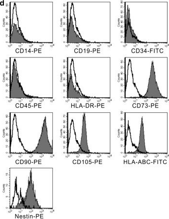
In Stem Cell Res Ther on 4 December 2014 by Wang, Y., Wu, H., et al.
Fig.2.D
-
FC/FACS
-
Homo sapiens (Human)
Collected and cropped from Stem Cell Res Ther by CiteAb, provided under a CC-BY license
Image 1 of 3
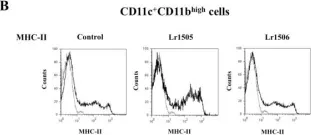
In BMC Immunol on 18 September 2012 by Villena, J., Chiba, E., et al.
Fig.8.B

-
FC/FACS
-
Collected and cropped from BMC Immunol by CiteAb, provided under a CC-BY license
Image 1 of 3
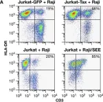
In PLoS Pathog on 26 February 2010 by Mazurov, D., Ilinskaya, A., et al.
Fig.6.A

-
FC/FACS
-
Homo sapiens (Human)
Collected and cropped from PLoS Pathog by CiteAb, provided under a CC-BY license
Image 1 of 3