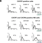
CXC Chemokine receptor type 4 (CXCR4) is commonly considered a potential marker for cancer stem cells (CSCs). Dedifferentiated-type chordoma (DTC) cells derived from a patient with recurrent chordoma exhibit high CXCR4 expression and demonstrate increased resistance to chemotherapeutic drugs and ionizing radiation (IR) compared to the conventional-type chordoma cell line, U-CH1. However, the precise role of CXCR4 in the stemness and IR resistance of DTC remains unclear. Therefore, this study aims to elucidate the correlation between the expression of CXCR4 and stemness and radioresistance in chordoma. DTC cells expressing CXCR4 (CXCR4+ DTC cells), isolated by magnetic-activated cell sorting, exhibited increased self-renewal activity, tumorigenicity, and IR resistance, accompanied by elevated Sox2 expression. Knockdown of CXCR4 expression using short hairpin RNA, inhibition of CXCR4 signaling with AMD3100, and targeting of STAT3, a downstream effector of CXCR4, with WP1066 in DTC cells significantly diminished their self-renewal ability, tumorigenic potential, IR resistance, and Sox2 expression. Additionally, transfection with a small interfering Sox2 RNA suppressed self-renewal activity, tumorigenicity, and IR resistance in DTC cells, whereas overexpression of CXCR4 reversed these effects in U-CH1 cells. Furthermore, DTC cells infected with shCXCR4 exhibited substantial tumor suppression, and the combination of IR and AMD3100 significantly reduced DTC tumor growth in a mouse xenograft model. These findings underscore the functional significance of CXCR4 as a CSC marker, highlighting its potential as a therapeutic target for malignant chordomas.
Product Citations: 148
CXCR4 confers stemness and radioresistance in chordoma cells.
In Cancer Biology & Therapy on 1 December 2025 by Jung, C. W., Kim, J. Y., et al.
-
Cancer Research
Preprint on BioRxiv : the Preprint Server for Biology on 16 April 2025 by Turner, D. L., Baric, H., et al.
The lung alveoli are constantly exposed to inhaled pathogens and inorganic hazards, relying on robust defence mechanisms to maintain homeostasis. Alveolar macrophages and type 2 alveolar epithelial cells (AT2s) collaborate to orchestrate protection. Compromised defence can dysregulate immunity and repair, leading to acute and chronic respiratory diseases. To better understand these processes and drive therapeutic discovery, human model systems that capture key cell interactions are essential. Here, we develop the first induced pluripotent stem cell (iPSC)-derived platform that integrates AT2 cells and macrophages in an air-liquid interface culture. Coculture enhanced AT2-specific gene expression and lipid synthesis, while macrophages actively phagocytosed AT2-derived surfactant. iPSC-derived AT2s supported macrophage survival by producing M-CSF and coculture promoted an alveolar macrophage-like phenotype. Additionally, during respiratory infection macrophages played a crucial role in modulating proinflammatory signalling, enhancing antiviral immunity, and restricting viral replication. Furthermore, we identify a role for iPSC-derived macrophages in epithelial repair, with VEGF signalling to macrophages increasing epithelial permeability. We present an iPSC-derived air-interface platform to study AT2-macrophage interactions in homeostasis, infection, and repair, providing insights into their potential roles in the initiation and progression of respiratory diseases.
-
Immunology and Microbiology
-
Stem Cells and Developmental Biology
In PLoS Pathogens on 1 June 2024 by Warner van Dijk, F. A., Tong, O., et al.
AXL+ Siglec-6+ dendritic cells (ASDC) are novel myeloid DCs which can be subdivided into CD11c+ and CD123+ expressing subsets. We showed for the first time that these two ASDC subsets are present in inflamed human anogenital tissues where HIV transmission occurs. Their presence in inflamed tissues was supported by single cell RNA analysis of public databases of such tissues including psoriasis diseased skin and colorectal cancer. Almost all previous studies have examined ASDCs as a combined population. Our data revealed that the two ASDC subsets differ markedly in their functions when compared with each other and to pDCs. Relative to their cell functions, both subsets of blood ASDCs but not pDCs expressed co-stimulatory and maturation markers which were more prevalent on CD11c+ ASDCs, thus inducing more T cell proliferation and activation than their CD123+ counterparts. There was also a significant polarisation of naïve T cells by both ASDC subsets toward Th2, Th9, Th22, Th17 and Treg but less toward a Th1 phenotype. Furthermore, we investigated the expression of chemokine receptors that facilitate ASDCs and pDCs migration from blood to inflamed tissues, their HIV binding receptors, and their interactions with HIV and CD4 T cells. For HIV infection, within 2 hours of HIV exposure, CD11c+ ASDCs showed a trend in more viral transfer to T cells than CD123+ ASDCs and pDCs for first phase transfer. However, for second phase transfer, CD123+ ASDCs showed a trend in transferring more HIV than CD11c+ ASDCs and there was no viral transfer from pDCs. As anogenital inflammation is a prerequisite for HIV transmission, strategies to inhibit ASDC recruitment into inflamed tissues and their ability to transmit HIV to CD4 T cells should be considered.
Copyright: © 2024 Warner van Dijk et al. This is an open access article distributed under the terms of the Creative Commons Attribution License, which permits unrestricted use, distribution, and reproduction in any medium, provided the original author and source are credited.
-
FC/FACS
-
Homo sapiens (Human)
-
Immunology and Microbiology
In Journal of Immunology Research on 8 May 2024 by Cevallos, C., Jarmoluk, P., et al.
This study aims to explore the influence of coinfection with HCV and HIV on hepatic fibrosis. A coculture system was set up to actively replicate both viruses, incorporating CD4 T lymphocytes (Jurkat), hepatic stellate cells (LX-2), and hepatocytes (Huh7.5). LX-2 cells' susceptibility to HIV infection was assessed through measurements of HIV receptor expression, exposure to cell-free virus, and cell-to-cell contact with HIV-infected Jurkat cells. The study evaluated profibrotic parameters, including programed cell death, ROS imbalance, cytokines (IL-6, TGF-β, and TNF-α), and extracellular matrix components (collagen, α-SMA, and MMP-9). The impact of HCV infection on LX-2/HIV-Jurkat was examined using soluble factors released from HCV-infected hepatocytes. Despite LX-2 cells being nonsusceptible to direct HIV infection, bystander effects were observed, leading to increased oxidative stress and dysregulated profibrotic cytokine release. Coculture with HIV-infected Jurkat cells intensified hepatic fibrosis, redox imbalance, expression of profibrotic cytokines, and extracellular matrix production. Conversely, HCV-infected Huh7.5 cells exhibited elevated profibrotic gene transcriptions but without measurable effects on the LX-2/HIV-Jurkat coculture. This study highlights how HIV-infected lymphocytes worsen hepatic fibrosis during HCV/HIV coinfection. They increase oxidative stress, profibrotic cytokine levels, and extracellular matrix production in hepatic stellate cells through direct contact and soluble factors. These insights offer valuable potential therapies for coinfected individuals.Copyright © 2024 Cintia Cevallos et al.
-
FC/FACS
-
Immunology and Microbiology
In American Journal of Human Genetics on 4 April 2024 by Jalil, S., Keskinen, T., et al.
Argininosuccinate lyase deficiency (ASLD) is a recessive metabolic disorder caused by variants in ASL. In an essential step in urea synthesis, ASL breaks down argininosuccinate (ASA), a pathognomonic ASLD biomarker. The severe disease forms lead to hyperammonemia, neurological injury, and even early death. The current treatments are unsatisfactory, involving a strict low-protein diet, arginine supplementation, nitrogen scavenging, and in some cases, liver transplantation. An unmet need exists for improved, efficient therapies. Here, we show the potential of a lipid nanoparticle-mediated CRISPR approach using adenine base editors (ABEs) for ASLD treatment. To model ASLD, we first generated human-induced pluripotent stem cells (hiPSCs) from biopsies of individuals homozygous for the Finnish founder variant (c.1153C>T [p.Arg385Cys]) and edited this variant using the ABE. We then differentiated the hiPSCs into hepatocyte-like cells that showed a 1,000-fold decrease in ASA levels compared to those of isogenic non-edited cells. Lastly, we tested three different FDA-approved lipid nanoparticle formulations to deliver the ABE-encoding RNA and the sgRNA targeting the ASL variant. This approach efficiently edited the ASL variant in fibroblasts with no apparent cell toxicity and minimal off-target effects. Further, the treatment resulted in a significant decrease in ASA, to levels of healthy donors, indicating restoration of the urea cycle. Our work describes a highly efficient approach to editing the disease-causing ASL variant and restoring the function of the urea cycle. This method relies on RNA delivered by lipid nanoparticles, which is compatible with clinical applications, improves its safety profile, and allows for scalable production.
Copyright © 2024 The Author(s). Published by Elsevier Inc. All rights reserved.
-
Genetics
In PLoS One on 24 August 2012 by Liberman, J., Sartelet, H., et al.
Fig.2.B

-
FC/FACS
-
Collected and cropped from PLoS One by CiteAb, provided under a CC-BY license
Image 1 of 1