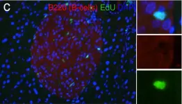
ALDH1L1 is widely used as a marker of astrocytes in the central nervous system (CNS), but its expression and potential roles in the periphery, particularly in lymphoid organs, remain poorly understood. Here, we found that ALDH1L1+ cells comprise approximately 5–9% of the lymph node (LN). To better understand their identity, we investigated whether these cells share characteristics with glial, immune, endothelial or stromal cell populations under homeostatic conditions. Using ALDH1L1/TdTomato reporter mice, immunofluorescence, and flow cytometry, we found that TdTomato+ cells in LNs do not coexpress canonical CNS (GFAP, ACSA-2) or peripheral glial (Sox10) markers. Similarly, using multiple approaches we found minimal overlap with T cells (CD3, TCRβ), B cells and dendritic cells (B220), myeloid cells (CD11b, Iba1), or antigen presenting cells (MHCII). To explore potential stromal associations, we assessed coexpression of TdTomato with endothelial (CD31, LYVE1) and reticular (ER-TR7) markers and found insubstantial overlap. In contrast, TdTomato+ cells showed significant colocalization with podoplanin (PDPN), a marker of fibroblastic reticular cells (FRCs). Interestingly, while many TdTomato+ cells were PDPN+, relatively few PDPN+ cells expressed TdTomato. Notably, ALDH1L1+PDPN+ cells were enriched in the paracortex and medulla but absent from the B cell follicles and subcapsular sinus. These findings suggest that while ALDH1L1 remains a reliable marker of astrocytes in the CNS, it may also delineate a distinct subset of FRCs in the LN, opening new avenues for exploring stromal cell diversity and function in lymphoid tissues.
Product Citations: 24
The astrocyte marker ALDH1L1 also identifies a stromal cell population in the lymph node.
In Scientific Reports on 9 February 2026 by Smith, B. C., Nasrallah, M. J., et al.
-
IHC-IF
-
Mus musculus (House mouse)
-
Neuroscience
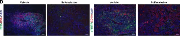
In Journal of the American Society of Nephrology : JASN on 1 February 2026 by Arai, H., Sugiura, Y., et al.
Tertiary lymphoid structure, an ectopic lymphoid tissue induced under chronic inflammation, develops in various kidney diseases and is associated with poor prognosis. The immune system requires metabolic resources to support immune function and lymphocyte proliferation. Hence, dramatic metabolic alterations presumably occur during the formation of tertiary lymphoid structure. However, it remains unclear whether metabolic remodeling occurs during this formation and its underlying mechanism.
In a murine model of renal tertiary lymphoid structures, we used imaging mass spectrometry and metabolome analysis to investigate the metabolic pathway that characterizes tertiary lymphoid structures. We also performed in situ hybridization with immunofluorescence and pharmacological inhibition to explore the expression and function of the key molecules governing the pivotal metabolic pathway. We analyzed urine samples from mice and humans to explore the metabolites estimating the presence of renal tertiary lymphoid structures.
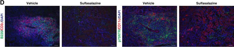
Significant glutathione accumulation and depletion of cysteine, which is essential for glutathione synthesis, was observed specifically within tertiary lymphoid structures. The kidneys with tertiary lymphoid structures exhibited higher glutathione concentrations than healthy kidneys. Tertiary lymphoid structures also showed significant accumulation of 4-HNE and 8-OHdG, markers of oxidative stress. Dendritic cells and fibroblasts within tertiary lymphoid structures expressed the cystine/glutamate transporter, that regulates glutathione synthesis, and supplied synthesized glutathione to lymphocytes, which lacked its expression. Pharmacological inhibition of the cystine/glutamate transporter prevented tertiary lymphoid structure formation in the kidney. Furthermore, enhanced glutathione synthesis within tertiary lymphoid structures was reflected in elevated urinary glutathione concentrations both in mice and humans, which effectively detected the presence of tertiary lymphoid structures in the kidney in IgA nephropathy patients.
Glutathione significantly accumulated within tertiary lymphoid structures in the kidney. Inhibition of the cystine/glutamate transporter prevented the formation of tertiary lymphoid structures. Urinary glutathione served as a biomarker to detect tertiary lymphoid structures in the kidney.
Copyright © 2025 The Author(s). Published by Wolters Kluwer Health, Inc. on behalf of the American Society of Nephrology.
-
IHC-IF
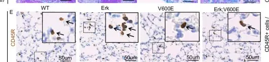
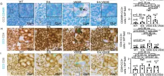
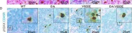
In Cell Death and Differentiation on 1 June 2024 by Bosso, G., Cintra Herpst, A. C., et al.
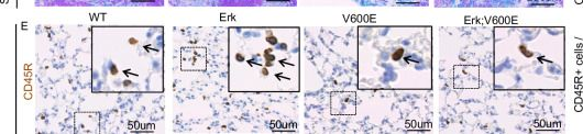
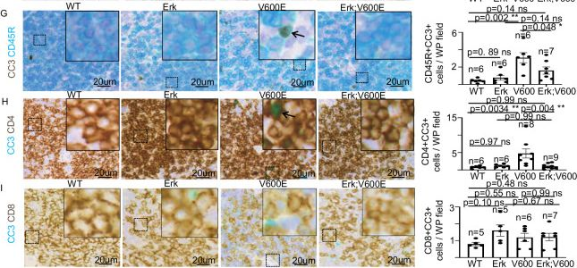
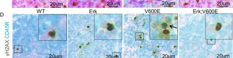
The BRAF gene is mutated in a plethora of human cancers. The majority of such molecular lesions result in the expression of a constitutively active BRAF variant (BRAFV600E) which continuously bolsters cell proliferation. Although we recently addressed the early effects triggered by BRAFV600E-activation, the specific contribution of ERK1 and ERK2 in BRAFV600E-driven responses in vivo has never been explored. Here we describe the first murine model suitable for genetically dissecting the ERK1/ERK2 impact in multiple phenotypes induced by ubiquitous BRAFV600E-expression. We unveil that ERK1 is dispensable for BRAFV600E-dependent lifespan shortening and for BRAFV600E-driven tumor growth. We show that BRAFV600E-expression provokes an ERK1-independent lymphocyte depletion which does not rely on p21CIP1-induced cell cycle arrest and is unresponsive to ERK-chemical inhibition. Moreover, we also reveal that ERK1 is dispensable for BRAFV600E-triggered cytotoxicity in lungs and that ERK-chemical inhibition abrogates some of these detrimental effects, such as DNA damage, in Club cells but not in pulmonary lymphocytes. Our data suggest that ERK1/ERK2 contribution to BRAFV600E-driven phenotypes is dynamic and varies dependently on cell type, the biological function, and the level of ERK-pathway activation. Our findings also provide useful insights into the comprehension of BRAFV600E-driven malignancies pathophysiology as well as the consequences in vivo of novel ERK pathway-targeted anti-cancer therapies.
© 2024. The Author(s).
-
IHC
-
WB
-
Mus musculus (House mouse)
IRF5 regulates microglial myelin clearance and cholesterol metabolism after demyelination
Preprint on BioRxiv : the Preprint Server for Biology on 16 August 2023 by Montilla, A., Zabala, A., et al.
Interferon regulatory factor 5 (IRF5), a transcription factor highly involved in innate immunity that drives microglia/macrophage towards a pro-inflammatory state, has been associated to multiple sclerosis susceptibility but its role in MS pathogenesis is unknown. Here we analysed the role of IRF5 in multiple sclerosis animal models. Irf 5 -/- mice showed exacerbated damage in the chronic phase of experimental autoimmune encephalomyelitis (EAE) mice, despite an initial delay in its onset, as well as after lysolecithin injection into the spinal cord. Transcriptomic and lipidomic analysis evidence a role of this transcription factor in myelin metabolism and cholesterol homeostasis. Indeed, Irf 5 -/- mice showed an aberrant accumulation of myelin debris and lipidic structures, such as CE-containing lipid droplets and cholesterol crystals, suggesting that myelin-derived lipids are not properly processed. Cholesterol crystal accumulation leads to an aberrant inflammatory response, which block oligodendrocyte migration into the core of demyelinated lesion and remyelination. Pharmacologically facilitating cholesterol transport reduces lipid droplet accumulation and ameliorates EAE exacerbated damage in Irf 5 -/- mice. These results reveal for the first time the role of Irf5, a transcription factor necessary to orchestrate the immune responses, in phagocytes lipid metabolism which could be pivotal in regenerative responses such as remyelination.
-
IHC
-
Mus musculus (House mouse)
-
Neuroscience
-
Cell Biology
-
Biochemistry and Molecular biology
In Cell Reports on 30 May 2023 by Bhin, J., Paes Dias, M., et al.
BRCA1 and BRCA2 both function in DNA double-strand break repair by homologous recombination (HR). Due to their HR defect, BRCA1/2-deficient cancers are sensitive to poly(ADP-ribose) polymerase inhibitors (PARPis), but they eventually acquire resistance. Preclinical studies yielded several PARPi resistance mechanisms that do not involve BRCA1/2 reactivation, but their relevance in the clinic remains elusive. To investigate which BRCA1/2-independent mechanisms drive spontaneous resistance in vivo, we combine molecular profiling with functional analysis of HR of matched PARPi-naive and PARPi-resistant mouse mammary tumors harboring large intragenic deletions that prevent reactivation of BRCA1/2. We observe restoration of HR in 62% of PARPi-resistant BRCA1-deficient tumors but none in the PARPi-resistant BRCA2-deficient tumors. Moreover, we find that 53BP1 loss is the prevalent resistance mechanism in HR-proficient BRCA1-deficient tumors, whereas resistance in BRCA2-deficient tumors is mainly induced by PARG loss. Furthermore, combined multi-omics analysis identifies additional genes and pathways potentially involved in modulating PARPi response.
Copyright © 2023 The Author(s). Published by Elsevier Inc. All rights reserved.
-
IHC
-
Mus musculus (House mouse)
-
Cancer Research
In J Am Soc Nephrol on 1 February 2026 by Arai, H., Sugiura, Y., et al.
Fig.4.D
-
IHC-IF
-
Collected and cropped from Journal of the American Society of Nephrology : JASN by CiteAb, provided under a CC-BY license
Image 1 of 5
In Cell Death Differ on 1 June 2024 by Bosso, G., Cintra Herpst, A. C., et al.
Fig.2.U
-
IHC
-
Mus musculus (House mouse)
Collected and cropped from Cell Death and Differentiation by CiteAb, provided under a CC-BY license
Image 1 of 5
In Cell Death Differ on 1 June 2024 by Bosso, G., Cintra Herpst, A. C., et al.
Fig.4.G
-
IHC
-
Mus musculus (House mouse)
Collected and cropped from Cell Death and Differentiation by CiteAb, provided under a CC-BY license
Image 1 of 5
In Cell Death Differ on 1 June 2024 by Bosso, G., Cintra Herpst, A. C., et al.
Fig.4.D
-
IHC
-
Mus musculus (House mouse)
Collected and cropped from Cell Death and Differentiation by CiteAb, provided under a CC-BY license
Image 1 of 5
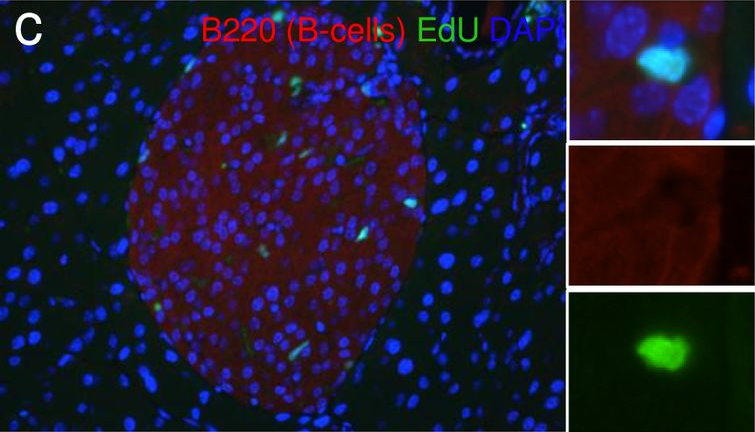
In PLoS One on 15 July 2016 by Cox, A. R., Barrandon, O., et al.
Fig.9.C
-
WB
-
Mus musculus (House mouse)
Collected and cropped from PLoS ONE by CiteAb, provided under a CC-BY license
Image 1 of 5