Prenatal stress is associated with a high risk of developing adult intestinal pathologies, such as irritable bowel syndrome, chronic inflammation, and cancer. Although epithelial stem cells and progenitors have been implicated in intestinal pathophysiology, how prenatal stress could impact their functions is still unknown. We have investigated the proliferative and differentiation capacities of primitive cells using epithelial crypts isolated from colons of adult male and female mice whose mothers have been stressed during late gestation. Our results show that stem cell/progenitor proliferation and differentiation in vitro are negatively impacted by prenatal stress in male progeny. This is promoted by a reinforcement of the negative proliferative/differentiation control by the protease-activated receptor 2 (PAR2) and the muscarinic receptor 3 (M3), two G protein-coupled receptors present in the crypt. Conversely, prenatal stress does not change in vitro proliferation of colon primitive cells in female progeny. Importantly, this maintenance is associated with a functional switch in the M3 negative control of colonoid growth, becoming proliferative after prenatal stress. In addition, the proliferative role of PAR2 specific to females is maintained under prenatal stress, even though PAR2-targeted stress signals Dusp6 and activated GSK3β are increased, reaching the levels of males. An epithelial serine protease could play a critical role in the activation of the survival kinase GSK3β in colonoids from prenatally stressed female progeny. Altogether, our results show that following prenatal stress, colon primitive cells cope with stress through sexually dimorphic mechanisms that could pave the way to dysregulated crypt regeneration and intestinal pathologies.NEW & NOTEWORTHY Primitive cells isolated from mouse colon following prenatal stress and exposed to additional stress conditions such as in vitro culture, present sexually dimorphic mechanisms based on PAR2- and M3-dependent regulation of proliferation and differentiation. Whereas prenatal stress reinforces the physiological negative control exerted by PAR2 and M3 in crypts from males, in females, it induces a switch in M3- and PAR2-dependent regulation leading to a resistant and proliferative phenotype of progenitor.
Product Citations: 67
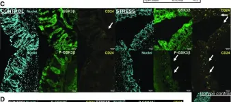
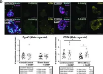
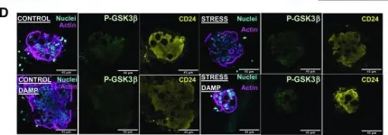
Prenatal stress induces changes in PAR2- and M3-dependent regulation of colon primitive cells.
In American Journal of Physiology - Gastrointestinal and Liver Physiology on 1 December 2022 by Berger, M., Guiraud, L., et al.
-
IHC-IF
-
ICC-IF
-
Mus musculus (House mouse)
-
Endocrinology and Physiology
Mesenchymal stromal cell apoptosis is required for their therapeutic function.
In Nature Communications on 11 November 2021 by Pang, S. H., D'Rozario, J., et al.
Multipotent mesenchymal stromal cells (MSCs) ameliorate a wide range of diseases in preclinical models, but the lack of clarity around their mechanisms of action has impeded their clinical utility. The therapeutic effects of MSCs are often attributed to bioactive molecules secreted by viable MSCs. However, we found that MSCs underwent apoptosis in the lung after intravenous administration, even in the absence of host cytotoxic or alloreactive cells. Deletion of the apoptotic effectors BAK and BAX prevented MSC death and attenuated their immunosuppressive effects in disease models used to define MSC potency. Mechanistically, apoptosis of MSCs and their efferocytosis induced changes in metabolic and inflammatory pathways in alveolar macrophages to effect immunosuppression and reduce disease severity. Our data reveal a mode of action whereby the host response to dying MSCs is key to their therapeutic effects; findings that have broad implications for the effective translation of cell-based therapies.
© 2021. The Author(s).
In Nature Communications on 4 March 2021 by Murray, M. P., Engel, I., et al.
Invariant natural killer T cells (iNKT cells) differentiate into thymic and peripheral NKT1, NKT2 and NKT17 subsets. Here we use RNA-seq and ATAC-seq analyses and show iNKT subsets are similar, regardless of tissue location. Lung iNKT cell subsets possess the most distinct location-specific features, shared with other innate lymphocytes in the lung, possibly consistent with increased activation. Following antigenic stimulation, iNKT cells undergo chromatin and transcriptional changes delineating two populations: one similar to follicular helper T cells and the other NK or effector like. Phenotypic analysis indicates these changes are observed long-term, suggesting that iNKT cells gene programs are not fixed, but they are capable of chromatin remodeling after antigen to give rise to additional subsets.
-
Immunology and Microbiology
In Blood Advances on 9 February 2021 by Paun, A., Claudio, E., et al.
There is a considerable body of work exploring the role of NF-κB family of transcription factors in the maturation and functions of later stage B cells; however, their role in the earlier bone marrow stages of development is less well understood despite the demonstration that NF-κB activity is present at all early stages of B-cell development. To explore the consequences of early, B cell-targeted constitutive activation of both NF-κB pathways on B-cell development, we generated mice that have either or both. NF-κB pathways constitutively activated beginning in early pro-B cells. In marked contrast to activating a single pathway, we found mice with both pathways constitutively activated displayed a profound loss of B cells, starting with early pro-B cells and peaking at the late pro-B-cell stage, at least in part as a result of increased apoptosis. This effect was found to be cell autonomous and to have striking phenotypic consequences on the secondary lymphoid organs and circulating antibody levels. This effect was also found to be temporal in nature as similar activation under a Cre expressed later in development did not result in generation of a similar phenotype. Taken together, these findings help to shed further light on the need for tight regulation of the NF-κB family of transcription factors during the various stages of B-cell development in the bone marrow.
-
Immunology and Microbiology
In The Journal of Immunology on 15 June 2020 by Meyer, S. J., Böser, A., et al.
B lymphocytes are important players of the adaptive immune system. However, not just activation of B cells but also regulation of B cell signaling is important to prevent hyperactivity and dysregulation of the immune response. Different mechanisms and proteins contribute to this balance. One of these is CD22, a member of the Siglec family. It is an inhibitory coreceptor of the BCR and inhibits B cell activation. Upon BCR stimulation, CD22-dependent inhibition of BCR signaling results in a decreased calcium mobilization. Although some CD22 binding partners have already been identified, the knowledge about the CD22 interactome is still incomplete. In this study, quantitative affinity purification-mass spectrometry enabled the delineation of the CD22 interactome in the B cell line DT40. These data will clarify molecular mechanisms and CD22 signaling events after BCR activation and revealed several new CD22-associated proteins. One new identified interaction partner is the E3 ubiquitin ligase cullin 3, which was revealed to regulate CD22 surface expression and clathrin-dependent CD22 internalization after BCR stimulation. Furthermore cullin 3 was identified to be important for B lymphocytes in general. B cell-specific cullin 3-deficient mice show reduced developing B cells in the bone marrow and a severe pro-B cell proliferation defect. Mature B cells in the periphery are also reduced and characterized by increased CD22 expression and additionally by preactivated and apoptotic phenotypes. The findings reveal novel functions of cullin 3 in B lymphocytes, namely regulating CD22 surface expression and internalization after B cell activation, as well as promoting proliferation of pro-B cells.
Copyright © 2020 by The American Association of Immunologists, Inc.
-
Immunology and Microbiology
In Am J Physiol Gastrointest Liver Physiol on 1 December 2022 by Berger, M., Guiraud, L., et al.
Fig.7.C

-
IHC-IF
-
Mus musculus (House mouse)
Collected and cropped from Am J Physiol Gastrointest Liver Physiol by CiteAb, provided under a CC-BY license
Image 1 of 6
In Am J Physiol Gastrointest Liver Physiol on 1 December 2022 by Berger, M., Guiraud, L., et al.
Fig.7.D

-
ICC-IF
-
Mus musculus (House mouse)
Collected and cropped from Am J Physiol Gastrointest Liver Physiol by CiteAb, provided under a CC-BY license
Image 1 of 6
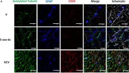
In Am J Physiol Gastrointest Liver Physiol on 1 December 2022 by Berger, M., Guiraud, L., et al.
Fig.8.D

-
IHC-IF
-
Mus musculus (House mouse)
Collected and cropped from Am J Physiol Gastrointest Liver Physiol by CiteAb, provided under a CC-BY license
Image 1 of 6
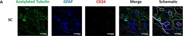
In Front Cell Dev Biol on 11 January 2020 by Rodríguez-Jiménez, F. J., Clemente, E., et al.
Fig.1.A

-
ICC
-
Mus musculus (House mouse)
Collected and cropped from Front Cell Dev Biol by CiteAb, provided under a CC-BY license
Image 1 of 6
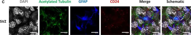
In Front Cell Dev Biol on 11 January 2020 by Rodríguez-Jiménez, F. J., Clemente, E., et al.
Fig.1.C

-
ICC
-
Mus musculus (House mouse)
Collected and cropped from Front Cell Dev Biol by CiteAb, provided under a CC-BY license
Image 1 of 6
In Front Cell Dev Biol on 11 January 2020 by Rodríguez-Jiménez, F. J., Clemente, E., et al.
Fig.6.A

-
ICC
-
Mus musculus (House mouse)
Collected and cropped from Front Cell Dev Biol by CiteAb, provided under a CC-BY license
Image 1 of 6