The persistence of chimeric antigen receptor (CAR) T cells in the tumor microenvironment limits their antitumor effects against solid tumors. Many studies have reported that the in vitro phenotype and metabolism of CAR-T cells correlates with their in vivo antitumor activity. Herein, we constructed PD-1 scFv-secreting and CD133-specific CAR-T (referred to as CAR-T) cells based on our previous work. We found that suitable concentration metformin-treated CAR-T (mCAR-T) cells exhibited an increased memory phenotype and demonstrated stronger and faster antitumor abilities with a reduced exhaustion phenotype. Using RNA sequencing, transmission electron microscope, and metabolic analysis, we discovered enhanced mitochondrial biogenesis and metabolism in CAR-T cells treated with 10 μM metformin, is associated with increased peroxisome proliferator-activated receptor gamma coactivator-1alpha (PGC-1α) expression, promotion of signal transducer and activator of transcription (STAT)3 and inhibition of STAT5 phosphorylation. This resulted in enhanced antitumor effects of mCAR-T cells in both subcutaneous and orthotopic xenograft models. Importantly, in some relapsed hepatocellular carcinoma (HCC) patients, high CD133 expression was observed in their paired primary or metastatic tumor sections. Our study revealed that enhancing metabolic fitness and central memory by in vitro metformin treatment is an effective strategy to improve the efficacy of CAR-T cell therapy, potentially benefiting some relapsed HCC patients.
© The author(s).
Product Citations: 108
In International Journal of Biological Sciences on 4 July 2025 by You, J., Yang, X., et al.
-
Biochemistry and Molecular biology
-
Cancer Research
-
Cell Biology
-
Immunology and Microbiology
In Bone Research on 24 February 2025 by Li, Z., Jiang, J., et al.
Knee arthrofibrosis, characterized by excessive matrix protein production and deposition, substantially impairs basic daily functions, causing considerable distress and financial burden. However, the underlying pathomechanisms remain unclear. Here, we characterized the heterogeneous cell populations and cellular pathways by combination of flow cytometry and single-cell RNA-seq analysis of synovial tissues from six patients with or without knee arthrofibrosis. Increased macrophages and fibroblasts were observed with decreased numbers of fibroblast-like synoviocytes, endothelial cells, vascular smooth muscle cells, and T cells in the arthrofibrosis group compared with negative controls. Notably, fibroblasts were discovered to interact with macrophages, and lead to fibrosis through TGF-β pathway induced CCN2 expression in fibroblasts. CCN2 was demonstrated to be required for fibroblast pro-fibrotic functions (activation, proliferation, and migration) through TGFBR/SMAD pathway. The expression of CCN2 was positively correlated with the collagen volume and TGF-β expression and negatively associated with patient-reported outcome measures in another cohort of patients with knee arthrofibrosis. Our study reveals the role of CCN2 in the fibroblast-macrophage interaction through TGF-β pathway which might help to shed light on CCN2 as a potential biomarker.
© 2025. The Author(s).
-
FC/FACS
-
Homo sapiens (Human)
-
Genetics
-
Immunology and Microbiology
In Journal of Hematology & Oncology on 18 December 2024 by Fang, H., Dai, W., et al.
Liver metastasis from colorectal cancer (CRC) is a major clinical challenge that severely affects patient survival. myofibroblastic cancer-associated fibroblasts (myCAFs) are a major component of the CRC tumor microenvironment, where they contribute to tumor progression and metastasis through exosomes.
Single-cell analysis highlighted a notable increase in myCAFs in colorectal cancer liver metastases (CRLM). Exosomal sequencing identified PWAR6 as the most significantly elevated lncRNA in these metastatic tissues. In vivo and in vitro assays confirmed PWAR6's roles in CRC cell stemness, migration, and glutamine uptake. RNA pulldown, RIP, and Co-IP assays investigated the molecular mechanisms of the PWAR6/NRF2/SLC38A2 signaling axis in CRC progression, flow cytometry was used to assess NK cell activity and cytotoxicity.
Clinically, higher PWAR6 expression levels are strongly associated with increased 68Ga FAPI-PET/CT SUVmax values, particularly in CRLM patients, where expression significantly exceeds that of non-LM cases and normal colon tissues. Regression analysis and survival data further support PWAR6 as a negative prognostic marker, with elevated levels correlating with worse patient outcomes. Mechanistically, PWAR6 promotes immune evasion by inhibiting NRF2 degradation through competitive binding with Keap1, thereby upregulating SLC38A2 expression, which enhances glutamine uptake in CRC cells and depletes glutamine availability for NK cells.
myCAFs derived exosomes PWAR6 represents a pivotal marker for CRC liver metastasis, and its targeted inhibition with ASO-PWAR6, in combination with FAPI treatment, effectively curtails metastasis in preclinical models, offering promising therapeutic potential for clinical management.
© 2024. The Author(s).
-
Cancer Research
CD28 Costimulation Augments CAR Signaling in NK Cells via the LCK/CD3ζ/ZAP70 Signaling Axis.
In Cancer Discovery on 4 October 2024 by Acharya, S., Basar, R., et al.
Multiple factors in the design of a chimeric antigen receptor (CAR) influence CAR T-cell activity, with costimulatory signals being a key component. Yet, the impact of costimulatory domains on the downstream signaling and subsequent functionality of CAR-engineered natural killer (NK) cells remains largely unexplored. Here, we evaluated the impact of various costimulatory domains on CAR-NK cell activity, using a CD70-targeting CAR. We found that CD28, a costimulatory molecule not inherently present in mature NK cells, significantly enhanced the antitumor efficacy and long-term cytotoxicity of CAR-NK cells both in vitro and in multiple xenograft models of hematologic and solid tumors. Mechanistically, we showed that CD28 linked to CD3ζ creates a platform that recruits critical kinases, such as lymphocyte-specific protein tyrosine kinase (LCK) and zeta-chain-associated protein kinase 70 (ZAP70), initiating a signaling cascade that enhances CAR-NK cell function. Our study provides insights into how CD28 costimulation enhances CAR-NK cell function and supports its incorporation in NK-based CARs for cancer immunotherapy. Significance: We demonstrated that incorporation of the T-cell-centric costimulatory molecule CD28, which is normally absent in mature natural killer (NK) cells, into the chimeric antigen receptor (CAR) construct recruits key kinases including lymphocyte-specific protein tyrosine kinase and zeta-chain-associated protein kinase 70 and results in enhanced CAR-NK cell persistence and sustained antitumor cytotoxicity.
©2024 American Association for Cancer Research.
-
Cancer Research
In Balkan Medical Journal on 6 September 2024 by Isildar, B., Ozkan, S., et al.
Mesenchymal stem cells (MSCs) play a key role in regenerative medicine due to their capacity to differentiate into multiple cell lines, regulate the immune system, and exert paracrine effects. The therapeutic impact of MSCs is primarily mediated through their secretome. The secretory and therapeutic potential of MSCs can be improved through preconditioning, which entails the application of hypoxic environments, 3-dimensional cell cultures, and pharmacological agents. Valproic acid (VPA) is a histone deacetylase inhibitor that is employed in medical practice for treating epilepsy and bipolar disorder. Hence, preconditioning MSCs with VPA is expected to induce histone acetylation, enhance gene expression, and beneficially modify the cells' secretomes.
To assess the effectiveness of VPA in enhancing and regulating the therapeutic potential of cells as well as its impact on MSC secretome profiles and ultrastructural morphologies.
Expiremental study.
Human umbilical cord MSCs were preconditioned with 2 mM VPA for 24 and 48 hours; untreated MSCs served as controls. The secretome secreted by the cells was assessed for its total protein content. Subsequently, interferon-gamma (IFN-γ), interleukin-17 (IL-17), IL-10, vascular endothelial growth factor, nerve growth factor (NGF), glial cell line-derived neurotrophic factor, and brain-derived neurotrophic factor (BDNF) levels in the secretome were analyzed using the ELISA method. The ultrastructural properties of the cells were studied under transmission electron microscopy.
Ultrastructural examinations revealed that the chromatin content of VPA-treated cells was reduced. VPA-preconditioned cells exhibited a higher density of rough endoplasmic reticulum, autophagic vesicles, and myelin figures on cytoplasmic structure analysis, which was indicative of increased secretion. Protein secretion was elevated in those cells, with notable increases in NGF and BDNF levels. Furthermore, the cytoskeletal rearrangement and elevated autophagic activity observed in the 48-hour preconditioned cells could indicate the initiation of neuronal differentiation. IL-10, IL-17, and IFN-γ were not detected in the secretome.
This study indicate that preconditioning with VPA enhances MSC activity and subsequently modifies the secretome content.
-
Homo sapiens (Human)
-
Genetics
-
Stem Cells and Developmental Biology
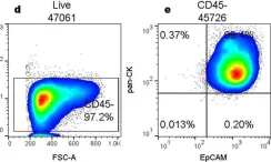
In Sci Rep on 6 February 2017 by Maestre-Batlle, D., Pena, O. M., et al.
Fig.1.D

-
FC/FACS
-
Homo sapiens (Human)
Collected and cropped from Sci Rep by CiteAb, provided under a CC-BY license
Image 1 of 1