The capacity of adipose stem/progenitor cells (ASCs) to undergo self-renewal and differentiation is crucial for adipose tissue homoeostasis, regeneration and expansion. However, the heterogeneous ASC populations of the adipose lineage constituting adipose tissue are not precisely known. In the present study, we demonstrate that cell surface expression of dipeptidyl peptidase-4 (DPP4)/cluster of differentiation 26 (CD26) subdivides the DLK1-/CD34+/CD45-/CD31- ASC pool of human white adipose tissues (WATs) into two large populations. Ex vivo, DPP4+ ASCs possess higher self-renewal and proliferation capacity and lesser adipocyte differentiation potential than DDP4- ASCs. The knock-down of DPP4 in ASC leads to significantly reduced proliferation and self-renewal capacity, while adipogenic differentiation is increased. Ectopic overexpression of DPP4 strongly inhibits adipogenesis. Moreover, in whole mount stainings of human subcutaneous (s)WAT, we detect DPP4 in CD34+ ASC located in the vascular stroma surrounding small blood vessels and in mature adipocytes. We conclude that DPP4 is a functional marker for an abundant ASC population in human WAT with high proliferation and self-renewal potential and low adipogenic differentiation capacity.
Product Citations: 11
In Adipocyte on 1 December 2022 by Hatzmann, F. M., Großmann, S., et al.
ChipCytometry for multiplexed detection of protein and mRNA markers on human FFPE tissue samples.
In STAR Protocols on 17 June 2022 by Jarosch, S., Köhlen, J., et al.
In this protocol, we describe the use of ChipCytometry to combine RNA in situ hybridization and antibody staining for multiplexed tissue imaging of human formalin-fixed and paraffin-embedded tissue samples. The advantages of ChipCytometry are long-term storage for re-interrogation and advanced image quality by high dynamic range imaging of staining and background. A titrated pretreatment of tissue samples bypasses challenges because of the retrieval of antigens on coverslips and achieves an optimal staining quality at the minimal expense of tissue integrity. For complete details on the use and execution of this protocol, please refer to Jarosch et al. (2021).
© 2022 The Author(s).
-
Genetics
Monocytes are the main source of STING-mediated IFN-α production.
In EBioMedicine on 1 June 2022 by Congy-Jolivet, N., Cenac, C., et al.
Type I interferon (IFN-I) production by plasmacytoid dendritic cells (pDCs) occurs during viral infection, in response to Toll-like receptor 7 (TLR7) stimulation and is more vigorous in females than in males. Whether this sex bias persists in ageing people is currently unknown. In this study, we investigated the effect of sex and aging on IFN-α production induced by PRR agonist ligands.
In a large cohort of individuals from 19 to 97 years old, we measured the production of IFN-α and inflammatory cytokines in whole-blood upon stimulation with either R-848, ODN M362 CpG-C, or cGAMP, which activate the TLR7/8, TLR9 or STING pathways, respectively. We further characterized the cellular sources of IFN-α.
We observed a female predominance in IFN-α production by pDCs in response to TLR7 or TLR9 ligands. The higher TLR7-driven IFN-α production in females was robustly maintained across ages, including the elderly. The sex-bias in TLR9-driven interferon production was lost after age 60, which correlated with the decline in circulating pDCs. By contrast, STING-driven IFN-α production was similar in both sexes, preserved with aging, and correlated with circulating monocyte numbers. Indeed, monocytes were the primary cellular source of IFN-α in response to cGAMP.
We show that the sex bias in the TLR7-induced IFN-I production is strongly maintained through ages, and identify monocytes as the main source of IFN-I production via STING pathway.
This work was supported by grants from Région Occitanie/Pyrénées-Méditerranée (#12052910, Inspire Program #1901175), University Paul Sabatier, and the European Regional Development Fund (MP0022856).
Copyright © 2022 The Authors. Published by Elsevier B.V. All rights reserved.
-
FC/FACS
-
Homo sapiens (Human)
In Cell Rep Methods on 22 November 2021 by Jarosch, S., Köhlen, J., et al.
Deciphering the spatial composition of cells in tissues is essential for detailed understanding of biological processes in health and disease. Recent technological advances enabled the assessment of the enormous complexity of tissue-derived parameters by highly multiplexed tissue imaging (HMTI), but elaborate machinery and data analyses are required. This severely limits broad applicability of HMTI. Here we demonstrate for the first time the application of ChipCytometry technology, which has unique features for widespread use, on formalin-fixed paraffin-embedded samples, the most commonly used storage technique of clinically relevant patient specimens worldwide. The excellent staining quality permits workflows for automated quantification of signal intensities, which we further optimized to compensate signal spillover from neighboring cells. In combination with the high number of validated markers, the reported platform can be used from unbiased analyses of tissue composition to detection of phenotypically complex rare cells, and can be easily implemented in both routine research and clinical pathology.
© 2021 The Authors.
-
Homo sapiens (Human)
In Cell Reports on 23 July 2019 by Ziegler, C. G. K., Kim, J., et al.
In cancer biology, the functional interpretation of genomic alterations is critical to achieve the promise of genomic profiling in the clinic. For chronic lymphocytic leukemia (CLL), a heterogeneous disease of B-lymphocytes maturing under constitutive B cell receptor (BCR) stimulation, the functional role of diverse clonal mutations remains largely unknown. Here, we demonstrate that alterations in BCR signaling dynamics underlie the progression of B cells toward malignancy. We reveal emergent dynamic features-bimodality, hypersensitivity, and hysteresis-in the BCR signaling pathway of primary CLL B cells. These signaling abnormalities in CLL quantitatively derive from BCR clustering and constitutive signaling with positive feedback reinforcement, as demonstrated through single-cell analysis of phospho-responses, computational modeling, and super-resolution imaging. Such dysregulated signaling segregates CLL patients by disease severity and clinical presentation. These findings provide a quantitative framework and methodology to assess complex and heterogeneous leukemia pathology and to inform therapeutic strategies in parallel with genomic profiling.
Copyright © 2019 The Author(s). Published by Elsevier Inc. All rights reserved.
-
Homo sapiens (Human)
-
Cancer Research
-
Immunology and Microbiology
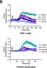
In Arthritis Res Ther on 13 July 2011 by Chang, B. Y., Huang, M. M., et al.
Fig.4.B

-
FC/FACS
-
Collected and cropped from Arthritis Res Ther by CiteAb, provided under a CC-BY license
Image 1 of 1