Oligodendrocyte progenitor cells (OPCs) represent a population of electrically active and dividing cells, which are capable of responding to neuronal activity by proliferating and differentiating. Here, we report that the repressive euchromatic H3K9me2 histone mark, deposited by the histone methyltransferases EHMT1 and EHMT2 enzymes, increases in proliferating OPCs in mice following optogenetic stimulation of neuronal activity. Using primary cultured OPCs with genetic deletion of Ehmt1 and Ehmt2, and pharmacological inhibition of EHMT enzymatic activity, we reveal the importance of these enzymes in repressing the expression of genes regulating cell death and electrical properties in proliferating OPCs. Consistent with these findings, we detect higher levels of cholinergic muscarinic receptors, fewer numbers of oligodendrocyte lineage cells, and lower levels of the myelin basic protein (MBP) in mice with lineage-specific ablation of Ehmt1 and Ehmt2. Together these data suggest that the repressive H3K9me2 histone mark, whose levels increase in proliferating OPCs after neuronal stimulation, is an important modulator of cell death and proteins regulating the electrical properties of OPCs.
© 2025 Wiley Periodicals LLC.
Product Citations: 219
In GLIA on 1 July 2025 by Pruvost, M., Park, H. J., et al.
-
Genetics
-
Neuroscience
In GLIA on 1 June 2025 by Wright, J. L., Jiang, Y., et al.
Chromatin remodeling complexes (CRCs) participate in oligodendrocyte (OL) differentiation, survival, and maintenance. We asked whether CRCs also control the proliferation of OL precursors (OPs)-focusing on the INO80 complex, which is known to regulate the proliferation of a variety of other cell types during development and disease. CRISPR/Cas9-mediated inactivation of Ino80 in vitro, or Cre-mediated deletion in vivo, slowed the OP cell cycle substantially by prolonging G1. RNAseq analysis revealed that E2F target genes were dysregulated in OPs from INO80-deficient mice, but correlated RNAseq and ATAC-seq uncovered no general correlation between gene expression and altered nucleosome positioning at transcription start sites. Fluorescence photobleaching experiments in cultured OPs demonstrated that histone H2A.Z mobility increased following the loss of INO80, suggesting that INO80 regulates the cell cycle machinery in OPs through H2A.Z/H2A exchange. We also present evidence that INO80 associates with OLIG2, a master regulator of OL development.
© 2025 The Author(s). GLIA published by Wiley Periodicals LLC.
-
Genetics
-
Neuroscience
Type I collagen secreted in white matter lesions inhibits remyelination and functional recovery.
In Cell Death & Disease on 13 April 2025 by Yamazaki, R., Azuma, M., et al.
White matter injury is caused by cerebral blood flow disturbances associated with stroke and demyelinating diseases such as multiple sclerosis. Remyelination is induced spontaneously after white matter injury, but progressive multiple sclerosis and white matter stroke are usually characterised by remyelination failure. However, the mechanisms underlying impaired remyelination in lesions caused by demyelination and stroke remain unclear. In the current study, we demonstrated that collagen fibres accumulated in the demyelinated lesions of multiple sclerosis patients (age range 23-80 years) and white matter lesions of stroke patients (age range 80-87 years), suggesting that the accumulation of collagen fibres correlates with remyelination failure in these lesions. To investigate the function of collagen fibres in the white matter lesions, we generated two types of white matter injury in mice. We induced focal demyelination by lysolecithin (LPC) injection and ischemic stroke by endothelin 1 (ET1) injection into the internal capsule. We found that type I collagen fibres were secreted in ET1-induced lesions with impaired white matter regeneration in the chronic phase of disease. We also showed that monocyte-derived macrophages that infiltrated into lesions from the peripheral blood produced type I collagen after white matter injury, and that type I collagen also exacerbated microglial activation, astrogliosis, and axonal injury. Finally, we demonstrated that oligodendrocyte differentiation and remyelination were inhibited in the presence of type I collagen after LPC-induced demyelination. These results suggest that type I collagen secreted by monocyte-derived macrophages inhibited white matter regeneration, and therefore, the modulation of type I collagen metabolism might be a novel therapeutic target for white matter injury.
© 2025. The Author(s).
-
IHC
-
Mus musculus (House mouse)
-
Cell Biology
In eLife on 14 March 2025 by Bose, M., Talwar, I., et al.
In the developing vertebrate central nervous system, neurons and glia typically arise sequentially from common progenitors. Here, we report that the transcription factor Forkhead Box G1 (Foxg1) regulates gliogenesis in the mouse neocortex via distinct cell-autonomous roles in progenitors and postmitotic neurons that regulate different aspects of the gliogenic FGF signalling pathway. We demonstrate that loss of Foxg1 in cortical progenitors at neurogenic stages causes premature astrogliogenesis. We identify a novel FOXG1 target, the pro-gliogenic FGF pathway component Fgfr3, which is suppressed by FOXG1 cell-autonomously to maintain neurogenesis. Furthermore, FOXG1 can also suppress premature astrogliogenesis triggered by the augmentation of FGF signalling. We identify a second novel function of FOXG1 in regulating the expression of gliogenic cues in newborn neocortical upper-layer neurons. Loss of FOXG1 in postmitotic neurons non-autonomously enhances gliogenesis in the progenitors via FGF signalling. These results fit well with the model that newborn neurons secrete cues that trigger progenitors to produce the next wave of cell types, astrocytes. If FGF signalling is attenuated in Foxg1 null progenitors, they progress to oligodendrocyte production. Therefore, loss of FOXG1 transitions the progenitor to a gliogenic state, producing either astrocytes or oligodendrocytes depending on FGF signalling levels. Our results uncover how FOXG1 integrates extrinsic signalling via the FGF pathway to regulate the sequential generation of neurons, astrocytes, and oligodendrocytes in the cerebral cortex.
© 2024, Bose et al.
In GLIA on 1 January 2025 by Liu, K., Kang, Z., et al.
Brain vasculature formation begins with vessel invasion from the perineural vascular plexus, which expands through vessel sprouting and growth. Recent studies have indicated the existence of oligodendrocyte-vascular crosstalk during development. However, the relationship between oligodendrocyte progenitor cells (OPCs) and the ordered spatiotemporal vascularization of the neocortex has not been elucidated. Our findings suggest that OPCs play a complex role in the vessel density of the embryonic and postnatal neocortex. Analyses of normal human and mouse embryonic cerebral cortex show that vascularization and OPC distribution are tightly controlled in a spatially and temporally restricted manner, exhibiting a positive correlation. Loss of OPCs at both embryonic and postnatal stages led to a reduction in vascular density, suggesting that OPC populations play a role in vascular density. Nonetheless, dynamic observation on cultured brain slices and staining of tissue sections indicate that OPC migration is unassociated with the proximity to blood vessels, primarily occurring along radial glial cell processes. Additionally, in vitro experiments demonstrate that OPC secretions promote vascular endothelial cell (VEC) growth. Together, these observations suggest that vessel density is influenced by OPC secretions.
© 2024 Wiley Periodicals LLC.
-
Mus musculus (House mouse)
-
Neuroscience
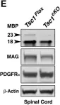
In Front Cell Neurosci on 28 July 2018 by Shi, Q., Saifetiarova, J., et al.
Fig.2.E

-
WB
-
Mus musculus (House mouse)
Collected and cropped from Front Cell Neurosci by CiteAb, provided under a CC-BY license
Image 1 of 1