Summary The genome of esophageal adenocarcinoma (EAC) is highly unstable and might evolve over time. Here, we track karyotype evolution in EACs in response to treatment and upon recurrence through multi-region and longitudinal analysis. To this end, we introduce L-PAC, a bio-informatics technique that allows inference of absolute copy number aberrations (CNA) of low-purity samples by leveraging information of high-purity samples from the same cancer. Quantitative analysis of matched absolute CNAs reveals that the amount of karyotype evolution induced by chemoradiotherapy (CRT) is predictive for early recurrence and depends on the initial level of karyotype intra-tumor heterogeneity. We observe that CNAs acquired in response to CRT are partially reversed back to the initial state upon recurrence. CRT hence alters the fitness landscape to which tumors can adjust by adapting their karyotype. Together, our results indicate that karyotype plasticity contributes to therapy resistance of EACs.
Product Citations: 29
Preprint on BioRxiv : the Preprint Server for Biology on 2 March 2024 by van der Sluis, K., van Sandick, J. W., et al.
-
Homo sapiens (Human)
Identification of ligand-receptor pairs that drive human astrocyte development.
In Nature Neuroscience on 1 August 2023 by Voss, A. J., Lanjewar, S., et al.
Extrinsic signaling between diverse cell types is crucial for nervous system development. Ligand binding is a key driver of developmental processes. Nevertheless, it remains a significant challenge to disentangle which and how extrinsic signals act cooperatively to affect changes in recipient cells. In the developing human brain, cortical progenitors transition from neurogenesis to gliogenesis in a stereotyped sequence that is in part influenced by extrinsic ligands. Here we used published transcriptomic data to identify and functionally test five ligand-receptor pairs that synergistically drive human astrogenesis. We validate the synergistic contributions of TGFβ2, NLGN1, TSLP, DKK1 and BMP4 ligands on astrocyte development in both hCOs and primary fetal tissue. We confirm that the cooperative capabilities of these five ligands are greater than their individual capacities. Additionally, we discovered that their combinatorial effects converge in part on the mTORC1 signaling pathway, resulting in transcriptomic and morphological features of astrocyte development. Our data-driven framework can leverage single-cell and bulk genomic data to generate and test functional hypotheses surrounding cell-cell communication regulating neurodevelopmental processes.
© 2023. The Author(s), under exclusive licence to Springer Nature America, Inc.
-
Neuroscience
Immunohistochemical characterization of bipolar cells in four distantly related avian species.
In Journal of Comparative Neurology on 1 March 2023 by Balaji, V., Haverkamp, S., et al.
Visual (and probably also magnetic) signal processing starts at the first synapse, at which photoreceptors contact different types of bipolar cells, thereby feeding information into different processing channels. In the chicken retina, 15 and 22 different bipolar cell types have been identified based on serial electron microscopy and single-cell transcriptomics, respectively. However, immunohistochemical markers for avian bipolar cells were only anecdotally described so far. Here, we systematically tested 12 antibodies for their ability to label individual bipolar cells in the bird retina and compared the eight most suitable antibodies across distantly related species, namely domestic chicken, domestic pigeon, common buzzard, and European robin, and across retinal regions. While two markers (GNB3 and EGFR) labeled specifically ON bipolar cells, most markers labeled in addition to bipolar cells also other cell types in the avian retina. Staining pattern of four markers (CD15, PKCα, PKCβ, secretagogin) was species-specific. Two markers (calbindin and secretagogin) showed a different expression pattern in central and peripheral retina. For the chicken and European robin, we found slightly more ON bipolar cell somata in the inner nuclear layer than OFF bipolar cell somata. In contrast, OFF bipolar cells made more ribbon synapses than ON bipolar cells in the inner plexiform layer of these species. Finally, we also analyzed the photoreceptor connectivity of selected bipolar cell types in the European robin retina. In summary, we provide a catalog of bipolar cell markers for different bird species, which will greatly facilitate analyzing the retinal circuitry of birds on a larger scale.
© 2022 The Authors. The Journal of Comparative Neurology published by Wiley Periodicals LLC.
-
Neuroscience
Developmental errors in the common marmoset retina.
In Frontiers in Neuroanatomy on 8 October 2022 by Haverkamp, S., Mietsch, M., et al.
Although retinal organization is remarkably conserved, morphological anomalies can be found to different extents and varieties across animal species with each presenting unique characteristics and patterns of displaced and misplaced neurons. One of the most widely used non-human primates in research, the common marmoset (Callithrix jaccus) could potentially also be of interest for visual research, but is unfortunately not well characterized in this regard. Therefore, the aim of our study was to provide a first time description of structural retinal layering including morphological differences and distinctive features in this species. Retinas from animals (n = 26) of both sexes and different ages were immunostained with cell specific antibodies to label a variety of bipolar, amacrine and ganglion cells. Misplaced ganglion cells with somata in the outermost part of the inner nuclear layer and rod bipolar cells with axon terminals projecting into the outer plexiform layer instead of the inner plexiform layer independent of age or sex of the animals were the most obvious findings, whereas misplaced amacrine cells and misplaced cone bipolar axon terminals occurred to a lesser extent. With this first time description of developmental retinal errors over a wide age range, we provide a basic characterization of the retinal system of the common marmosets, which can be taken into account for future studies in this and other animal species. The finding of misplaced ganglion cells and misplaced bipolar cell axon terminals was not reported before and displays an anatomic variation worthwhile for future analyzes of their physiological and functional impact.
Copyright © 2022 Haverkamp, Mietsch and Briggman.
-
Stem Cells and Developmental Biology
No evidence for age-related alterations in the marmoset retina.
In Frontiers in Neuroanatomy on 20 September 2022 by Haverkamp, S., Reinhard, K., et al.
The physiological aging process of the retina is accompanied by various and sometimes extensive changes: Macular degeneration, retinopathies and glaucoma are the most common findings in the elderly and can potentially lead to irreversible visual disablements up to blindness. To study the aging process and to identify possible therapeutic targets to counteract these diseases, the use of appropriate animal models is mandatory. Besides the most commonly used rodent species, a non-human primate, the common marmoset (Callithrix jacchus) emerged as a promising animal model of human aging over the last years. However, the visual aging process in this species is only partially characterized, especially with regard to retinal aberrations. Therefore, we assessed here for the first time potential changes in retinal morphology of the common marmoset of different age groups. By cell type specific immunolabeling, we analyzed different cell types and distributions, potential photoreceptor and ganglion cell loss, and structural reorganization. We detected no signs of age-related differences in staining patterns or densities of various cell populations. For example, there were no signs of photoreceptor degeneration, and there was only minimal sprouting of rod bipolar cells in aged retinas. Altogether, we describe here the maintenance of a stable neuronal architecture, distribution and number of different cell populations with only mild aberrations during the aging process in the common marmoset retina. These findings are in stark contrast to previously reported findings in rodent species and humans and deserve further investigations to identify the underlying mechanisms and possible therapeutic targets.
Copyright © 2022 Haverkamp, Reinhard, Peichl and Mietsch.
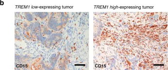
In Sci Rep on 1 November 2017 by Saurer, L., Zysset, D., et al.
Fig.5.B

-
IHC
-
Collected and cropped from Sci Rep by CiteAb, provided under a CC-BY license
Image 1 of 1