Age-related macular degeneration (AMD) is a complex multifactorial degenerative disease that leads to irreversible blindness. AMD affects the macula, the central part of the retina responsible for sharp central vision. Retinal pigment epithelium (RPE) is the main cellular type affected in dry AMD. RPE cells form a monolayer between the choroid and the neuroretina and are in close functional relationship with photoreceptors; moreover, RPE cells are part of the blood retina barrier that is disrupted in ocular diseases such as AMD. During ocular inflammation lymphocytes and macrophages are recruited, contact RPE and produce pro-inflammatory cytokines, which play an important role in AMD pathogenesis. The interaction between RPE and immune cells is mediated by leukocyte integrins, heterodimeric transmembrane receptors, and adhesion molecules, including VCAM-1 and ICAM-1. Within this frame, this study aimed to characterize RPE-leukocytes interaction and to investigate any potentially beneficial effects induced by integrin antagonists (DS-70, MN27 and SR714), developed in previous studies. ARPE-19 cells were co-cultured for different incubation times with Jurkat cells and apoptosis and necrosis levels were analyzed by flow cytometry. Moreover, we measured the mRNA levels of the pro-inflammatory cytokine IL-1β and the expression of adhesion molecules VCAM-1 and ICAM-1. We found that RPE-lymphocyte interaction increased apoptosis and necrosis levels in RPE cells and the expression of IL-1β. This interaction was mediated by the binding of α4β1 and αLβ2 integrins to VCAM-1 and ICAM-1, respectively. The blockade of RPE-lymphocyte interaction with blocking antibodies highlighted the pivotal role played by integrins. Therefore, α4β1 and αLβ2 integrin antagonists were employed to disrupt RPE-lymphocyte crosstalk. Small molecule integrin antagonists proved to be effective in reducing RPE cell death and expression of IL-1β, demonstrating that integrin antagonists could protect RPE cells from detrimental effects induced by the interaction with immune cells recruited to the retina. Overall, the leukocyte integrin antagonists employed in the present study may represent a novel opportunity to develop new drugs to fight dry AMD.
Copyright © 2021 Baiula, Caligiana, Bedini, Zhao, Santino, Cirillo, Gentilucci, Giacomini and Spampinato.
Product Citations: 13
Leukocyte Integrin Antagonists as a Novel Option to Treat Dry Age-Related Macular Degeneration.
In Frontiers in Pharmacology on 16 February 2021 by Baiula, M., Caligiana, A., et al.
-
Pharmacology
In Cell Host & Microbe on 8 May 2019 by Assil, S., Coléon, S., et al.
Type I interferon (IFN-I) is critical for antiviral defense, and plasmacytoid dendritic cells (pDCs) are a predominant source of IFN-I during virus infection. pDC-mediated antiviral responses are stimulated upon physical contact with infected cells, during which immunostimulatory viral RNA is transferred to pDCs, leading to IFN production via the nucleic acid sensor TLR7. Using dengue, hepatitis C, and Zika viruses, we demonstrate that the contact site of pDCs with infected cells is a specialized platform we term the interferogenic synapse, which enables viral RNA transfer and antiviral responses. This synapse is formed via αLβ2 integrin-ICAM-1 adhesion complexes and the recruitment of the actin network and endocytic machinery. TLR7 signaling in pDCs promotes interferogenic synapse establishment and provides feed-forward regulation, sustaining pDC contacts with infected cells. This interferogenic synapse may allow pDCs to scan infected cells and locally secrete IFN-I, thereby confining a potentially deleterious response.
Copyright © 2019 Elsevier Inc. All rights reserved.
-
Immunology and Microbiology
-
Neuroscience
In Mediators of Inflammation on 14 June 2016 by Ashander, L. M., Appukuttan, B., et al.
Targeting the endothelial adhesion molecules that control leukocyte trafficking into a tissue has been explored as a biological therapy for inflammatory diseases. However, these molecules also participate in leukocyte migration for immune surveillance, and inhibiting the physiological level of an adhesion molecule might promote infection or malignancy. We explored the concept of targeting endothelial adhesion molecule transcription during inflammation in a human system. Intercellular adhesion molecule 1 (ICAM-1) mediates leukocyte migration across the retinal endothelium in noninfectious posterior uveitis. We observed an increase in the transcription factor, nuclear factor of kappa light polypeptide gene enhancer in B-cells 1 (NF-κB1), in parallel with ICAM-1, in human retinal endothelial cells treated with tumor necrosis factor-alpha (TNF-α), and identified putative binding sites for NF-κB1 within the ICAM-1 regulatory region. We targeted induced NF-κB1 expression in endothelial cells with small interfering (si)RNA. Knockdown of NF-κB1 significantly decreased cell surface expression of ICAM-1 protein induced by TNF-α but did not reduce constitutive ICAM-1 expression. Consistently, NF-κB1 knockdown significantly reduced leukocyte binding to cell monolayers in the presence of TNF-α but did not impact baseline binding. Findings of this proof-of-concept study indicate that induced transcription of endothelial adhesion molecules might be targeted therapeutically for inflammatory disease in humans.
-
Biochemistry and Molecular biology
-
Immunology and Microbiology
Basic motifs target PSGL-1, CD43, and CD44 to plasma membrane sites where HIV-1 assembles.
In Journal of Virology on 1 January 2015 by Grover, J. R., Veatch, S. L., et al.
HIV-1 incorporates various host membrane proteins during particle assembly at the plasma membrane; however, the mechanisms mediating this incorporation process remain poorly understood. We previously showed that the HIV-1 structural protein Gag localizes to the uropod, a rear-end structure of polarized T cells, and that assembling Gag copatches with a subset, but not all, of the uropod-directed proteins, i.e., PSGL-1, CD43, and CD44, in nonpolarized T cells. The latter observation suggests the presence of a mechanism promoting virion incorporation of these cellular proteins. To address this possibility and identify molecular determinants, in the present study we examined coclustering between Gag and the transmembrane proteins in T and HeLa cells using quantitative two-color superresolution localization microscopy. Consistent with the findings of the T-cell copatching study, we found that basic residues within the matrix domain of Gag are required for Gag-PSGL-1 coclustering. Notably, the presence of a polybasic sequence in the PSGL-1 cytoplasmic domain significantly enhanced this coclustering. We also found that polybasic motifs present in the cytoplasmic tails of CD43 and CD44 also promote their coclustering with Gag. ICAM-1 and ICAM-3, uropod-directed proteins that do not copatch with Gag in T cells, and CD46, a non-uropod-directed protein, showed no or little coclustering with Gag. However, replacing their cytoplasmic tails with the cytoplasmic tail of PSGL-1 significantly enhanced their coclustering with Gag. Altogether, these results identify a novel mechanism for host membrane protein association with assembling HIV-1 Gag in which polybasic sequences present in the cytoplasmic tails of the membrane proteins and in Gag are the major determinants.
Nascent HIV-1 particles incorporate many host plasma membrane proteins during assembly. However, it is largely unknown what mechanisms promote the association of these proteins with virus assembly sites within the plasma membrane. Notably, our previous study showed that HIV-1 structural protein Gag colocalizes with a group of uropod-directed transmembrane proteins, PSGL-1, CD43, and CD44, at the plasma membrane of T cells. The results obtained in the current study using superresolution localization microscopy suggest the presence of a novel molecular mechanism promoting the association of PSGL-1, CD43, and CD44 with assembling HIV-1 which relies on polybasic sequences in HIV-1 Gag and in cytoplasmic domains of the transmembrane proteins. This information advances our understanding of virion incorporation of host plasma membrane proteins, some of which modulate virus spread positively or negatively, and suggests a possible new strategy to enrich HIV-1-based lentiviral vectors with a desired transmembrane protein.
Copyright © 2015, American Society for Microbiology. All Rights Reserved.
-
Immunology and Microbiology
In PLoS ONE on 1 February 2012 by Pan, D., Das, A., et al.
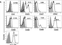
Impairment of intestinal epithelial barriers contributes to the progression of HIV/SIV infection and leads to generalized HIV-induced immune-cell activation during chronic infection. Rhesus macaques are the major animal model for studying HIV pathogenesis. However, detailed characterization of isolated rhesus epithelial cells (ECs) from intestinal tissues is not well defined. It is also not well documented whether isolated ECs had any other cell contaminants from intestinal tissues during the time of processing that might hamper interpretation of EC preparations or cultures. In this study, we identify and characterize ECs based on flow cytometry and immunohistochemistry methods using various enzymatic and mechanical isolation techniques to enrich ECs from intestinal tissues. This study shows that normal healthy ECs differentially express HLA-DR, CD23, CD27, CD90, CD95 and IL-10R markers. Early apoptosis and upregulation of ICAM-1 and HLA-DR in intestinal ECs are thought to be the key features in SIV mediated enteropathy. The data suggest that intestinal ECs might be playing an important role in mucosal immune responses by regulating the expression of different important regulatory and adhesion molecules and their function.
-
FC/FACS
In PLoS One on 1 February 2012 by Pan, D., Das, A., et al.
Fig.8.A

-
FC/FACS
-
Collected and cropped from PLoS One by CiteAb, provided under a CC-BY license
Image 1 of 1