Production and emigration of neural crest cells is a transient process followed by the emergence of the definitive roof plate. The mechanisms regulating the end of neural crest ontogeny are poorly understood. Whereas early crest development is stimulated by mesoderm-derived retinoic acid, we report that the end of the neural crest period is regulated by retinoic acid synthesized in the dorsal neural tube. Inhibition of retinoic acid signaling in the neural tube prevents the normal upregulation of BMP inhibitors in the nascent roof plate and prolongs the period of BMP responsiveness which otherwise ceases close to roof plate establishment. Consequently, neural crest production and emigration are extended well into the roof plate stage. In turn, extending the activity of neural crest-specific genes inhibits the onset of retinoic acid synthesis in roof plate suggesting a mutual repressive interaction between neural crest and roof plate traits. Although several roof plate-specific genes are normally expressed in the absence of retinoic acid signaling, roof plate and crest markers are co-expressed in single cells and this domain also contains dorsal interneurons. Hence, the cellular and molecular architecture of the roof plate is compromised. Collectively, our results demonstrate that neural tube-derived retinoic acid, via inhibition of BMP signaling, is an essential factor responsible for the end of neural crest generation and the proper segregation of dorsal neural lineages.
© 2022, Rekler and Kalcheim.
Product Citations: 8
In eLife on 8 April 2022 by Rekler, D. & Kalcheim, C.
-
IHC
Tissue-Specific Regulation of HNK-1 Biosynthesis by Bisecting GlcNAc.
In Molecules (Basel, Switzerland) on 26 August 2021 by Kawade, H., Morise, J., et al.
Human natural killer-1 (HNK-1) is a sulfated glyco-epitope regulating cell adhesion and synaptic functions. HNK-1 and its non-sulfated forms, which are specifically expressed in the brain and the kidney, respectively, are distinctly biosynthesized by two homologous glycosyltransferases: GlcAT-P in the brain and GlcAT-S in the kidney. However, it is largely unclear how the activity of these isozymes is regulated in vivo. We recently found that bisecting GlcNAc, a branching sugar in N-glycan, suppresses both GlcAT-P activity and HNK-1 expression in the brain. Here, we observed that the expression of non-sulfated HNK-1 in the kidney is unexpectedly unaltered in mutant mice lacking bisecting GlcNAc. This suggests that the biosynthesis of HNK-1 in the brain and the kidney are differentially regulated by bisecting GlcNAc. Mechanistically, in vitro activity assays demonstrated that bisecting GlcNAc inhibits the activity of GlcAT-P but not that of GlcAT-S. Furthermore, molecular dynamics simulation showed that GlcAT-P binds poorly to bisected N-glycan substrates, whereas GlcAT-S binds similarly to bisected and non-bisected N-glycans. These findings revealed the difference of the highly homologous isozymes for HNK-1 synthesis, highlighting the novel mechanism of the tissue-specific regulation of HNK-1 synthesis by bisecting GlcNAc.
Bisecting GlcNAc Is a General Suppressor of Terminal Modification of N-glycan.
In Molecular & Cellular Proteomics : MCP on 1 October 2019 by Nakano, M., Mishra, S. K., et al.
Glycoproteins are decorated with complex glycans for protein functions. However, regulation mechanisms of complex glycan biosynthesis are largely unclear. Here we found that bisecting GlcNAc, a branching sugar residue in N-glycan, suppresses the biosynthesis of various types of terminal epitopes in N-glycans, including fucose, sialic acid and human natural killer-1. Expression of these epitopes in N-glycan was elevated in mice lacking the biosynthetic enzyme of bisecting GlcNAc, GnT-III, and was conversely suppressed by GnT-III overexpression in cells. Many glycosyltransferases for N-glycan terminals were revealed to prefer a nonbisected N-glycan as a substrate to its bisected counterpart, whereas no up-regulation of their mRNAs was found. This indicates that the elevated expression of the terminal N-glycan epitopes in GnT-III-deficient mice is attributed to the substrate specificity of the biosynthetic enzymes. Molecular dynamics simulations further confirmed that nonbisected glycans were preferentially accepted by those glycosyltransferases. These findings unveil a new regulation mechanism of protein N-glycosylation.
© 2019 Nakano et al.
-
Biochemistry and Molecular biology
Perineuronal Net Protein Neurocan Inhibits NCAM/EphA3 Repellent Signaling in GABAergic Interneurons.
In Scientific Reports on 18 April 2018 by Sullivan, C. S., Gotthard, I., et al.
Perineuronal nets (PNNs) are implicated in closure of critical periods of synaptic plasticity in the brain, but the molecular mechanisms by which PNNs regulate synapse development are obscure. A receptor complex of NCAM and EphA3 mediates postnatal remodeling of inhibitory perisomatic synapses of GABAergic interneurons onto pyramidal cells in the mouse frontal cortex necessary for excitatory/inhibitory balance. Here it is shown that enzymatic removal of PNN glycosaminoglycan chains decreased the density of GABAergic perisomatic synapses in mouse organotypic cortical slice cultures. Neurocan, a key component of PNNs, was expressed in postnatal frontal cortex in apposition to perisomatic synapses of parvalbumin-positive interneurons. Polysialylated NCAM (PSA-NCAM), which is required for ephrin-dependent synapse remodeling, bound less efficiently to neurocan than mature, non-PSA-NCAM. Neurocan bound the non-polysialylated form of NCAM at the EphA3 binding site within the immunoglobulin-2 domain. Neurocan inhibited NCAM/EphA3 association, membrane clustering of NCAM/EphA3 in cortical interneuron axons, EphA3 kinase activation, and ephrin-A5-induced growth cone collapse. These studies delineate a novel mechanism wherein neurocan inhibits NCAM/EphA3 signaling and axonal repulsion, which may terminate postnatal remodeling of interneuron axons to stabilize perisomatic synapses in vivo.
In Scientific Reports on 21 September 2017 by Rice, R., Cebra-Thomas, J., et al.
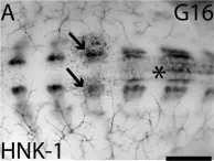
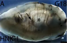
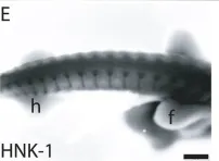
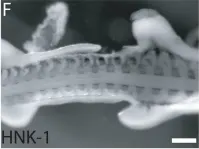
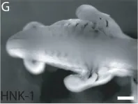
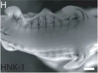
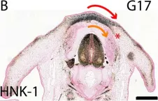
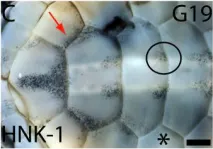
Ectothermal reptiles have internal pigmentation, which is not seen in endothermal birds and mammals. Here we show that the development of the dorsal neural tube-derived melanoblasts in turtle Trachemys scripta is regulated by similar mechanisms as in other amniotes, but significantly later in development, during the second phase of turtle trunk neural crest emigration. The development of melanoblasts coincided with a morphological change in the dorsal neural tube between stages mature G15 and G16. The melanoblasts delaminated and gathered in the carapacial staging area above the neural tube at G16, and differentiated into pigment-forming melanocytes during in vitro culture. The Mitf-positive melanoblasts were not restricted to the dorsolateral pathway as in birds and mammals but were also present medially through the somites similarly to ectothermal anamniotes. This matched a lack of environmental barrier dorsal and lateral to neural tube and the somites that is normally formed by PNA-binding proteins that block entry to medial pathways. PNA-binding proteins may also participate in the patterning of the carapacial pigmentation as both the migratory neural crest cells and pigment localized only to PNA-free areas.
-
IHC
-
IHC-P
-
Trachemys scripta elegans (Red-eared slider)
In Sci Rep on 21 September 2017 by Rice, R., Cebra-Thomas, J., et al.
Fig.6.A

-
IHC-P
-
Trachemys scripta elegans (Red-eared slider)
Collected and cropped from Sci Rep by CiteAb, provided under a CC-BY license
Image 1 of 8
In Sci Rep on 21 September 2017 by Rice, R., Cebra-Thomas, J., et al.
Fig.7.A

-
IHC-P
-
Trachemys scripta elegans (Red-eared slider)
Collected and cropped from Sci Rep by CiteAb, provided under a CC-BY license
Image 1 of 8
In Sci Rep on 21 September 2017 by Rice, R., Cebra-Thomas, J., et al.
Fig.2.E

-
IHC-P
-
Trachemys scripta elegans (Red-eared slider)
Collected and cropped from Sci Rep by CiteAb, provided under a CC-BY license
Image 1 of 8
In Sci Rep on 21 September 2017 by Rice, R., Cebra-Thomas, J., et al.
Fig.2.F

-
IHC-P
-
Trachemys scripta elegans (Red-eared slider)
Collected and cropped from Sci Rep by CiteAb, provided under a CC-BY license
Image 1 of 8
In Sci Rep on 21 September 2017 by Rice, R., Cebra-Thomas, J., et al.
Fig.2.G

-
IHC-P
-
Trachemys scripta elegans (Red-eared slider)
Collected and cropped from Sci Rep by CiteAb, provided under a CC-BY license
Image 1 of 8
In Sci Rep on 21 September 2017 by Rice, R., Cebra-Thomas, J., et al.
Fig.2.H

-
IHC-P
-
Trachemys scripta elegans (Red-eared slider)
Collected and cropped from Sci Rep by CiteAb, provided under a CC-BY license
Image 1 of 8
In Sci Rep on 21 September 2017 by Rice, R., Cebra-Thomas, J., et al.
Fig.6.B

-
IHC-P
-
Trachemys scripta elegans (Red-eared slider)
Collected and cropped from Sci Rep by CiteAb, provided under a CC-BY license
Image 1 of 8
In Sci Rep on 21 September 2017 by Rice, R., Cebra-Thomas, J., et al.
Fig.7.C

-
IHC-P
-
Trachemys scripta elegans (Red-eared slider)
Collected and cropped from Sci Rep by CiteAb, provided under a CC-BY license
Image 1 of 8
