
The majority of the eukaryotic cell surface is decorated with a layer of membrane-attached polysaccharides and glycoproteins collectively referred to as the glycocalyx. While the formation of a bulky glycocalyx has been associated with the cancer progression, the mechanisms by which the glycocalyx regulates cancer invasiveness are incompletely understood. We address this question by first documenting subtype-specific expression of the major glycocalyx glycoprotein Mucin-1 (MUC1) in breast cancer patient samples and breast cancer cell lines. Strikingly, glycocalyx disruption led to inhibition of 2D motility, loss of 3D invasion, and reduction of clonal scattering in breast cancer cells at the population level. Tracking of 2D cell motility and 3D invasiveness of MUC1-based sorted subpopulations revealed the fastest motility and invasiveness in intermediate MUC1-expressing cells, with glycocalyx disruption abolishing these effects. While differential sensitivity in 2D motility is attributed to a nonmonotonic dependence of focal adhesion size on MUC1 levels, higher MUC1 levels enhance 3D invasiveness via increased traction generation. In contrast to inducing cell rounding on collagen-coated substrates, high MUC1 level promotes cell adhesion and confers resistance to shear flow on substrates coated with the endothelial surface protein E-selectin. Collectively, our findings illustrate how MUC1 drives cancer invasiveness by differentially regulating cell-substrate adhesion in a substrate-dependent manner.
© The Author(s) 2024. Published by Oxford University Press on behalf of National Academy of Sciences.
Product Citations: 37
Bulky glycocalyx drives cancer invasiveness by modulating substrate-specific adhesion.
In PNAS Nexus on 1 August 2024 by Barai, A., Piplani, N., et al.
-
Homo sapiens (Human)
-
Cancer Research
In Cancers on 20 May 2024 by Kriebardis, A. G., Chardalias, L., et al.
The release of microvesicles (MVs) is an essential phenomenon for inter-cellular signaling in health and disease. The role of MVs in cancer is multidimensional and includes cancer cell survival, proliferation, and invasion. In this prospective study, we analyzed MV levels in colorectal cancer patients and assessed the importance of MV release in early-stage colorectal cancer and survival.
This study included 98 patients and 15 controls. The characterization of MVs from human plasma was performed by flow cytometry using monoclonal antibodies.
The levels of total MVs and MUC-1-positive, tissue factor (TF)-positive, and endothelial cell-derived MVs (EMVs) were statistically significantly higher in the colon cancer patients than in the controls (p < 0.001). Furthermore, the subgroup of patients with very early-stage colorectal cancer also had statistically significant differences in the levels of the abovementioned MVs compared to the controls (p < 0.01). Highly differentiated tumors had lower levels of MUC-1-positive MVs (p < 0.02), EMVs (p < 0.002), and EMV/TF combinations (p < 0.001) versus those with tumors with low/intermediate differentiation.
Our data demonstrate that the analysis of circulating MV levels in plasma could possibly become a tool for the early diagnosis of colon cancer at a very early stage of the disease.
-
Homo sapiens (Human)
-
Cancer Research
In Nature Communications on 30 March 2024 by Giudice, E., Huang, T. T., et al.
The multi-cohort phase 2 trial NCT02203513 was designed to evaluate the clinical activity of the CHK1 inhibitor (CHK1i) prexasertib in patients with breast or ovarian cancer. Here we report the activity of CHK1i in platinum-resistant high-grade serous ovarian carcinoma (HGSOC) with measurable and biopsiable disease (cohort 5), or without biopsiable disease (cohort 6). The primary endpoint was objective response rate (ORR). Secondary outcomes were safety and progression-free survival (PFS). 49 heavily pretreated patients were enrolled (24 in cohort 5, 25 in cohort 6). Among the 39 RECISTv1.1-evaluable patients, ORR was 33.3% in cohort 5 and 28.6% in cohort 6. Primary endpoint was not evaluable due to early stop of the trial. The median PFS was 4 months in cohort 5 and 6 months in cohort 6. Toxicity was manageable. Translational research was an exploratory endpoint. Potential biomarkers were investigated using pre-treatment fresh biopsies and serial blood samples. Transcriptomic analysis revealed high levels of DNA replication-related genes (POLA1, POLE, GINS3) associated with lack of clinical benefit [defined post-hoc as PFS < 6 months]. Subsequent preclinical experiments demonstrated significant cytotoxicity of POLA1 silencing in combination with CHK1i in platinum-resistant HGSOC cell line models. Therefore, POLA1 expression may be predictive for CHK1i resistance, and the concurrent POLA1 inhibition may improve the efficacy of CHK1i monotherapy in this hard-to-treat population, deserving further investigation.
© 2024. This is a U.S. Government work and not under copyright protection in the US; foreign copyright protection may apply.
-
Cancer Research
Bulky glycocalyx drives cancer invasiveness by modulating substrate-specific adhesion
Preprint on BioRxiv : the Preprint Server for Biology on 6 August 2023 by Barai, A., Piplani, N., et al.
Majority of the eukaryotic cell surface is decorated with a layer of membrane attached polysaccharides and glycoproteins collectively referred to as the glycocalyx. While formation of a bulky glycocalyx has been associated with cancer progression, the mechanisms by which the glycocalyx regulates cancer invasiveness is incompletely understood. We address this question by first documenting sub-type specific expression of the major glycocalyx glycoprotein Mucin-1 (MUC1) in breast cancer patient samples and breast cancer cell lines. Strikingly, glycocalyx disruption led to inhibition of 2D motility, loss of 3D invasion and reduction of clonal scattering of breast cancer cells at the population level. Tracking of 2D cell motility and 3D invasiveness of MUC1-based sorted sub-populations revealed fastest motility and invasiveness in intermediate MUC1-expressing cells, with glycocalyx disruption abolishing these effects. While differential sensitivity in 2D motility is attributed to a non-monotonic dependence of focal adhesion size on MUC1 levels, higher MUC1 levels enhance 3D invasiveness via increased traction generation. In contrast to inducing cell rounding on collagen-coated substrates, high MUC1 level promotes cell adhesion and confers resistance to shear flow on substrates coated with the endothelial surface protein E-selectin. Collectively, our findings illustrate how MUC1 drives cancer invasiveness by differentially regulating cell-substrate adhesion in a substrate-dependent manner.
-
Cancer Research
Nintedanib induces apoptosis in human pterygium cells through the FGFR2-ERK signalling pathway.
In International Journal of Ophthalmology on 20 April 2023 by Gong, Y., Liao, Y. H., et al.
To investigate whether nintedanib can inhibit pterygium cells through the fibroblast growth factor receptor 2 (FGFR2)/extracellular-signal-regulated kinase (ERK) pathway.
Human primary pterygium cells were cultured in vitro. After treatment with nintedanib, the cell morphology was observed under microscopy, the morphological changes of the nucleus were observed after DAPI staining, apoptosis was analyzed by Annexin-V FITC/PI double staining, and the changes of apoptosis-associated proteins were detected by Western blot. The binding ability of nintedanib to FGFR2 was predicted by molecular docking. Finally, by silencing FGFR2, we explored whether nintedanib inhibited FGFR2/ERK pathway.
The results showed that nintedanib inhibited the growth of pterygium cells and caused nuclear pyknosis. The results of Annexin-VFITC/PI double staining showed that nintedanib was able to induce early and late apoptosis of pterygium cells, significantly increasing the expression of apoptosis-associated proteins Bax and cleaved-Caspase3 (P<0.05), and reducing the expression of Bcl-2 (P<0.05). In addition, nintedanib significantly inhibited ERK1/2 phosphorylation through FGFR2 (P<0.05). After silencing the expression of FGFR2, there was no significant difference in the inhibition of ERK1/2 phosphorylation by nintedanib (P>0.05).
Nintedanib induces apoptosis of pterygium cells by inhibiting FGFR2/ERK pathway.
International Journal of Ophthalmology Press.
-
FC/FACS
-
Homo sapiens (Human)
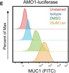
In Nat Commun on 15 July 2022 by Ferguson, I. D., Patiño-Escobar, B., et al.
Fig.5.E

-
FC/FACS
-
Collected and cropped from Nat Commun by CiteAb, provided under a CC-BY license
Image 1 of 3
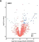
In Nat Commun on 15 July 2022 by Ferguson, I. D., Patiño-Escobar, B., et al.
Fig.5.C

-
FC/FACS
-
Collected and cropped from Nat Commun by CiteAb, provided under a CC-BY license
Image 1 of 3
In Nat Commun on 15 July 2022 by Ferguson, I. D., Patiño-Escobar, B., et al.
Fig.5.D

-
FC/FACS
-
Collected and cropped from Nat Commun by CiteAb, provided under a CC-BY license
Image 1 of 3