The BCR comprises a membrane-bound Ig that is noncovalently associated with a heterodimer of CD79A and CD79B. While the BCR Ig component functions to sense extracellular Ag, CD79 subunits contain cytoplasmic ITAMs that mediate intracellular propagation of BCR signals critical for B cell development, survival, and Ag-induced activation. CD79 is therefore an attractive target for Ab and chimeric Ag receptor T cell therapies for autoimmunity and B cell neoplasia. Although the mouse is an attractive model for preclinical testing, due to its well-defined immune system, an obstacle is the lack of cross-reactivity of candidate therapeutic anti-human mAbs with mouse CD79. To overcome this problem, we generated knockin mice in which the extracellular Ig-like domains of CD79A and CD79B were replaced with human equivalents. In this study, we describe the generation and characterization of mice expressing chimeric CD79 and report studies that demonstrate their utility in preclinical analysis of anti-human CD79 therapy. We demonstrate that human and mouse CD79 extracellular domains are functionally interchangeable, and that anti-human CD79 lacking Fc region effector function does not cause significant B cell depletion, but induces 1) decreased expression of plasma membrane-associated IgM and IgD, 2) uncoupling of BCR-induced tyrosine phosphorylation and calcium mobilization, and 3) increased expression of PTEN, consistent with the levels observed in anergic B cells. Finally, anti-human CD79 treatment prevents disease development in two mouse models of autoimmunity. We also present evidence that anti-human CD79 treatment may inhibit Ab secretion by terminally differentiated plasmablasts and plasma cells in vitro.Copyright © 2022 by The American Association of Immunologists, Inc.
Product Citations: 11
In The Journal of Immunology on 1 April 2022 by Wemlinger, S. M., Parker Harp, C. R., et al.
-
Immunology and Microbiology
In Immunity on 12 October 2021 by Sharma, M. D., Pacholczyk, R., et al.
Monocytic-lineage inflammatory Ly6c+CD103+ dendritic cells (DCs) promote antitumor immunity, but these DCs are infrequent in tumors, even upon chemotherapy. Here, we examined how targeting pathways that inhibit the differentiation of inflammatory myeloid cells affect antitumor immunity. Pharmacologic inhibition of Bruton's tyrosine kinase (BTK) and the tryptophan-degrading enzyme indoleamine 2,3-dioxygenase (IDO) or deletion of Btk or Ido1 allowed robust differentiation of inflammatory Ly6c+CD103+ DCs during chemotherapy, promoting antitumor T cell responses and inhibiting tumor growth. Immature Ly6c+c-kit+ precursor cells had epigenetic profiles similar to conventional DC precursors; deletion of Btk or Ido1 promoted differentiation of these cells. Mechanistically, a BTK-IDO axis inhibited a tryptophan-sensitive differentiation pathway driven by GATOR2 and mTORC1, and disruption of the GATOR2 in monocyte-lineage precursors prevented differentiation into inflammatory DCs in vivo. IDO-expressing DCs and monocytic cells were present across a range of human tumors. Thus, a BTK-IDO axis represses differentiation of inflammatory DCs during chemotherapy, with implications for targeted therapies.
Copyright © 2021 Elsevier Inc. All rights reserved.
-
Cancer Research
-
Immunology and Microbiology
In Frontiers in Immunology on 28 August 2020 by Agazio, A., Cimons, J., et al.
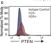
Peripheral tolerance is essential for silencing weakly autoreactive B cells that have escaped central tolerance, but it is unclear why these potentially pathogenic B cells are retained rather than being eliminated entirely. Release from peripheral tolerance restraint can occur under certain circumstances (i.e., strong TLR stimulus), that are present during infection. In this regard, we hypothesized that autoreactive B cells could function as a reserve population that can be activated to contribute to the humoral immune response, particularly with pathogens, such as HIV-1, that exploit immune tolerance to avoid host defense. In this study, we identify a population of autoreactive B cells with the potential to neutralize HIV-1 and experimentally release them from the functional restrictions of peripheral tolerance. We have previously identified murine monoclonal antibodies that displayed autoreactivity against histone H2A and neutralized HIV-1 in vitro. Here, we identify additional H2A-reactive IgM monoclonal antibodies and demonstrate that they are both autoreactive and polyreactive with self and foreign antigens and are able to neutralize multiple clades of tier 2 HIV-1. Flow cytometric analysis of H2A-reactive B cells in naïve wildtype mice revealed that these B cells are present in peripheral B cell populations and we further document that murine H2A-reactive B cells are restrained by peripheral tolerance mechanisms. Specifically, we show endogenous H2A-reactive B cells display increased expression of the inhibitory mediators CD5 and phosphatase and tensin homolog (PTEN) phosphatase and fail to mobilize calcium upon immunoreceptor stimulation; all characterized markers of anergy. Moreover, we show that toll-like receptor stimulation or provision of CD4 T cell help induces the in vitro production of H2A-reactive antibodies, breaking tolerance. Thus, we have identified a novel poly/autoreactive B cell population that has the potential to neutralize HIV-1 but is silenced by immune tolerance.
Copyright © 2020 Agazio, Cimons, Shotts, Guo, Santiago, Pelanda and Torres.
-
FC/FACS
-
Genetics
-
Immunology and Microbiology
In Frontiers in Oncology on 12 July 2019 by Giglio, S., Annibali, V., et al.
Endometrial cancer is the most common gynecologic malignancy in developed countries. Estrogen-dependent tumors (type I, endometrioid) account for 80% of cases and non-estrogen-dependent (type II, non-endometrioid) account for the rest. Endometrial cancer type I is generally thought to develop via precursor lesions along with the increasing accumulation of molecular genetic alterations. Endometrial hyperplasia with atypia/Endometrial Intraepithelial Neoplasia is the least common type of hyperplasia but it is the type most likely to progress to type I cancer, whereas endometrial hyperplasia without atypia rarely progresses to carcinoma. MicroRNAs are a class of small, non-coding, single-stranded RNAs that negatively regulate gene expression mainly binding to 3'-untranslated region of target mRNAs. In the current study, we identified a microRNAs signature (miR-205, miR-146a, miR-1260b) able to discriminate between atypical and typical endometrial hyperplasia in two independent cohorts of patients. The identification of molecular markers that can distinguish between these two distinct pathological conditions is considered to be highly useful for the clinical management of patients because hyperplasia with an atypical change is associated with a higher risk of developing cancer. We show that the combination of miR-205, -146a, and -1260b has the best predictive power in discriminating these two conditions (>90%). With the aim to find a biological role for these three microRNAs, we focused our attention on a common putative target involved in endometrial carcinogenesis: the oncosuppressor gene SMAD4. We showed that miRs-146a,-205, and-1260b directly target SMAD4 and their enforced expression induced proliferation and migration of Endometrioid Cancer derived cell lines, Hec1a cells. These data suggest that microRNAs-mediated impairment of the TGF-β pathway, due to inhibition of its effector molecule SMAD4, is a relevant molecular alteration in endometrial carcinoma development. Our findings show a potential diagnostic role of this microRNAs signature for the accurate diagnosis of Endometrial hyperplasia with atypia/Endometrial Intraepithelial Neoplasia and improve the understanding of their pivotal role in SMAD4 regulation.
-
WB
-
Homo sapiens (Human)
-
Genetics
Lymphocyte Activation Gene-3 Maintains Mitochondrial and Metabolic Quiescence in Naive CD4+ T Cells.
In Cell Reports on 2 April 2019 by Previte, D. M., Martins, C. P., et al.
Lymphocyte activation gene-3 (LAG-3) is an inhibitory receptor expressed by CD4+ T cells and tempers their homeostatic expansion. Because CD4+ T cell proliferation is tightly coupled to bioenergetics, we investigate the role of LAG-3 in modulating naive CD4+ T cell metabolism. LAG-3 deficiency enhances the metabolic profile of naive CD4+ T cells by elevating levels of mitochondrial biogenesis. In vivo, LAG-3 blockade partially restores expansion and the metabolic phenotype of wild-type CD4+ T cells to levels of Lag3-/- CD4+ T cells, solidifying that LAG-3 controls these processes. Lag3-/- CD4+ T cells also demonstrate greater signal transducer and activator of transcription 5 (STAT5) activation, enabling resistance to interleukin-7 (IL-7) deprivation. These results implicate this pathway as a target of LAG-3-mediated inhibition. Additionally, enhancement of STAT5 activation, as a result of LAG-3 deficiency, contributes to greater activation potential in these cells. These results identify an additional mode of regulation elicited by LAG-3 in controlling CD4+ T cell responses.
Copyright © 2019 The Author(s). Published by Elsevier Inc. All rights reserved.
-
Biochemistry and Molecular biology
-
Cell Biology
-
Immunology and Microbiology
In Front Immunol on 28 August 2020 by Agazio, A., Cimons, J., et al.
Fig.5.C

-
FC/FACS
-
Collected and cropped from Front Immunol by CiteAb, provided under a CC-BY license
Image 1 of 1