CD8+ T cells play an important role in anti-tumor immunity. Better understanding of their regulation could advance cancer immunotherapies. Here we identify, via stepwise CRISPR-based screening, that CUL5 is a negative regulator of the core signaling pathways of CD8+ T cells. Knocking out CUL5 in mouse CD8+ T cells significantly improves their tumor growth inhibiting ability, with significant proteomic alterations that broadly enhance TCR and cytokine signaling and their effector functions. Chemical inhibition of neddylation required by CUL5 activation, also enhances CD8 effector activities with CUL5 validated as a major target. Mechanistically, CUL5, which is upregulated by TCR stimulation, interacts with the SOCS-box-containing protein PCMTD2 and inhibits TCR and IL2 signaling. Additionally, CTLA4 is markedly upregulated by CUL5 knockout, and its inactivation further enhances the anti-tumor effect of CUL5 KO. These results together reveal a negative regulatory mechanism for CD8+ T cells and have strong translational implications in cancer immunotherapy.
© 2024. The Author(s).
Product Citations: 7
The CUL5 E3 ligase complex negatively regulates central signaling pathways in CD8+ T cells.
In Nature Communications on 19 January 2024 by Liao, X., Li, W., et al.
-
FC/FACS
-
Mus musculus (House mouse)
-
Immunology and Microbiology
In The Journal of Immunology on 15 October 2021 by Liang, Z., Zhang, Q., et al.
Thymic epithelial cells (TECs) are critical for the development and generation of functionally competent T cells. Until now, the mechanism that regulates the survival of TECs is poorly understood. In the current study, we found that Tsc1 controls the homeostasis of medullary TECs (mTECs) by inhibiting lysosomal-mediated apoptosis pathway in mice. TEC-specific deletion of Tsc1 predominately decreased the cell number of mTECs and, to a lesser content, affected the development cortical TECs. The defect of mTECs caused by Tsc1 deficiency in mice impaired thymocyte development and peripheral T cell homeostasis. Mechanistically, Tsc1 deficiency did not affect the cell proliferation of mTECs but increased the apoptosis of mTECs significantly. RNA-sequencing analysis showed that pathways involved in lysosomal biogenesis, cell metabolism, and apoptosis were remarkably elevated in Tsc1-deficient mTECs compared with their wild-type counterparts. Tsc1-deficient mTECs exhibited overproduction of reactive oxygen species and malfunction of lysosome, with lysosome membrane permeabilization and the release of cathepsin B and cathepsin L to the cytosol, which then lead to Bid cleaved into active truncated Bid and subsequently intrinsic apoptosis. Finally, we showed that the impaired development of mTECs could be partially reversed by decreasing mTORC1 activity via haploinsufficiency of Raptor Thus, Tsc1 is essential for the homeostasis of mTECs by inhibiting lysosomal-mediated apoptosis through mTORC1-dependent pathways.
Copyright © 2021 by The American Association of Immunologists, Inc.
-
Immunology and Microbiology
In Advanced Therapeutics on 1 August 2021 by Nam, J., Son, S., et al.
Photothermal therapy (PTT) and neoantigen cancer vaccine each offers minimally invasive and highly specific cancer therapy; however, they are not effective against large established tumors due to physical and biological barriers that attenuate thermal ablation and abolish anti-tumor immunity. Here, we designed and performed comparative study using small (~ 50 mm3) and large (> 100 mm3) tumors to examine how tumor size affects the therapeutic efficiency of PTT and neoantigen cancer vaccine. We show that spiky gold nanoparticle (SGNP)-based PTT and synergistic dual adjuvant-based neoantigen cancer vaccine can efficiently regress small tumors as a single agent, but not large tumors due to limited internal heating and immunosuppressive tumor microenvironment (TME). We report that PTT sensitizes tumors to neoantigen cancer vaccination by destroying and compromising the TME via thermally induced cellular and molecular damage, while neoantigen cancer vaccine reverts local immune suppression induced by PTT and shapes residual TME in favor of anti-tumor immunity. The combination therapy efficiently eradicated large local tumors and also exerted strong abscopal effect against pre-established distant tumors with robust systemic anti-tumor immunity. Thus, PTT combined with neoantigen cancer vaccine is a promising nano-immunotherapy for personalized therapy of advanced cancer.
-
Cancer Research
-
Immunology and Microbiology
Proinflammatory S100A8 Induces PD-L1 Expression in Macrophages, Mediating Tumor Immune Escape.
In The Journal of Immunology on 1 May 2020 by Li, Z., Wang, J., et al.
S100A8 is a damage-associated molecular pattern protein released by monocytes, playing a decisive role in the development of inflammation. Nonresolving inflammation is viewed as a driving force in tumorigenesis, and its role in tumor immune escape also attracted attentions. PD-1/PD-L1 axis is a critical determinant of physiological immune homeostasis, and anti-PD-1 or PD-L1 therapy has becoming the most exciting field of oncology. Multiple regulation mechanisms have been contributed to PD-L1 expression modulation including inflammatory mediators. In this study we reported that S100A8 significantly induced PD-L1 expression in monocytes/macrophages but not in tumor cells. S100A8 induced PD-L1 transcription through the TLR4 receptor and multiple crucial pathways of inflammation process. S100A8 modulated the histone modification of the PD-L1 promoter in monocytes/macrophages. S100A8-pretreated macrophages had immunosuppressive function and attenuated the antitumor ability of CTLs both in vitro and in vivo. A highly positive correlation existed between S100A8 expression and PD-L1 expression in human cancer specimens. To our knowledge, our study uncovers a novel molecular mechanism for regulating PD-L1 transcription by an inflammatory mediator S100A8, and reveals the importance of comprehensive understanding the role of inflammation in tumorigenesis as well as in tumor immune escape.
Copyright © 2020 by The American Association of Immunologists, Inc.
-
Cancer Research
-
Immunology and Microbiology
In Frontiers in Pharmacology on 6 April 2019 by Hansel, C., Erschfeld, S., et al.
Infiltrating CD4 and CD8 T cells have been shown to worsen inflammatory liver damage in non-alcoholic steatohepatitis (NASH). Inhibitory T cell receptors such as the programmed cell death protein 1 (PD1) and the natural killer cell receptor 2B4 regulate the activity of CD4 and CD8 T cells and therefore play an important role in immune tolerance required in the liver. In this study, we investigated the expression profile of inhibitory T cell receptors on CD4 and CD8 T cells in a mouse model of NASH. Male B57BL/6J mice were fed a Western diet for 24 weeks. The expression levels of inhibitory receptors on the surface of intrahepatic and peripheral T cells were measured and correlated with markers of activation (CD107a, CD69, and CD44), metabolic disorder (serum triglycerides, serum cholesterol, γ-glutamyl transferase, hepatic triglycerides), inflammation (serum alanine aminotransferase and aspartate aminotransferase) and hepatic fibrosis (collagen 1A1, α-smooth muscle actin, hydroxyproline). Under Western diet, PD1 is exclusively upregulated on intrahepatic and peripheral CD8+ T cells, whereas the expression level on CD4 T cells is unaffected. In contrast, 2B4 is upregulated liver-specifically on both CD4 and CD8 T cells and unchanged on peripheral T cells. Upregulation of PD1 on CD8 T cells is restricted to CD8 effector memory T cells and correlates with lower levels of degranulation. Similarly, the inhibitory function of PD1 on intrahepatic CD4 T cells is shown by a lower CD69 and CD44 expression on PD1-positive CD4 T cells. In murine steatohepatitis, the upregulation of PD1 on CD8 T cells and 2B4 on CD4 and CD8 T cells potentially limits T cell-mediated liver damage. Therefore, these inhibitory T cell receptors could serve as promising targets of immune-modulatory NASH therapy.
-
FC/FACS
-
Mus musculus (House mouse)
-
Immunology and Microbiology
-
Pharmacology
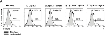
In PLoS One on 18 July 2015 by Silva, O., Crocetti, J., et al.
Fig.5.A

-
FC/FACS
-
Mus musculus (House mouse)
Collected and cropped from PLoS One by CiteAb, provided under a CC-BY license
Image 1 of 1