Tumor infiltrating lymphocytes (TIL) have demonstrated efficacious clinical outcomes for many patients with various types of solid cancers, including melanoma, gastrointestinal cancer, lung cancer, and head and neck cancer. Currently, the majority of clinical trials require that patients did not receive systemic therapy right before tumor tissue resection to avoid the interference of chemotherapy in the ex vivo TIL expansion. The primary disadvantage of this strategy is limiting the accessibility of TIL therapy for many eligible cancer patients. Over the past decade, substantial progress has been made for ex vivo expansion technologies in T cells. In this study, we investigated the possibility of enrolling patients who underwent chemotherapy prior to surgical resection. We collected seventeen tumor tissues from treatment naive cases, and five from cases that underwent chemotherapies. Cancer indications enrolled in this study were colorectal and lung cancers from both primary and metastatic sites, such as liver and brain. TILs from these tumors were expanded ex vivo to 2.1E8 (total viable lymphocytes counts) on average, with an overall success rate of 90.9%. Subsequently, TIL phenotypes and cytokine production were analyzed using flow cytometry and ELISA, respectively. We demonstrated functional TIL expansion from tumor tissues despite chemotherapy prior to surgical resection. We observed no significant phenotypic or functional differences between groups with and without chemotherapy. TIL expansion rate and characteristics were similar regardless of chemotherapy prior to resection, thereby providing a possibility to recruit patients with the most recent chemotherapy history in TIL therapy trials.
© 2023. The Author(s), under exclusive licence to Springer-Verlag GmbH Germany, part of Springer Nature.
Product Citations: 8
In Cancer Immunology, Immunotherapy : CII on 1 October 2023 by Balzeau, J., Ravindran, A., et al.
-
Cancer Research
Ad26.COV2.S priming provided a solid immunological base for mRNA-based COVID-19 booster vaccination.
In IScience on 20 January 2023 by Geers, D., Sablerolles, R. S. G., et al.
The emergence of novel SARS-CoV-2 variants led to the recommendation of booster vaccinations after Ad26.COV2.S priming. It was previously shown that heterologous booster vaccination induces high antibody levels, but how heterologous boosters affect other functional aspects of the immune response remained unknown. Here, we performed immunological profiling of Ad26.COV2.S-primed individuals before and after homologous or heterologous (mRNA-1273 or BNT162b2) booster. Booster vaccinations increased functional antibodies targeting ancestral SARS-CoV-2 and emerging variants. Especially heterologous booster vaccinations induced high levels of functional antibodies. In contrast, T-cell responses were similar in magnitude following homologous or heterologous booster vaccination and retained cross-reactivity towards variants. Booster vaccination led to a minimal expansion of SARS-CoV-2-specific T-cell clones and no increase in the breadth of the T-cell repertoire. In conclusion, we show that Ad26.COV2.S priming vaccination provided a solid immunological base for heterologous boosting, increasing humoral and cellular responses targeting emerging variants of concern.
© 2022 The Author(s).
-
COVID-19
-
Genetics
-
Immunology and Microbiology
In BMC Complementary Medicine and Therapies on 9 April 2021 by Olwenyi, O. A., Asingura, B., et al.
In Sub-Saharan Africa, herbal therapy continues to be utilized for HIV-1 disease management. However, the therapeutic benefits of these substances remain ambiguous. To date, little is known about the effects of these plant extracts on chronic CD4 + T-cell activation and exhaustion which is partly driven by HIV-1 associated microbial translocation.
Effects of Azadirachta indica, Momordica foetida and Moringa oleifera ethanol: water mixtures on cell viability were evaluated using the Guava PCA system. Then, an in-vitro cell culture model was developed to mimic CD4+ T cell exposures to antigens following HIV-1 microbial translocation. In this, peripheral blood mononuclear cells (PBMCs) isolated from HIV negative (n = 13), viral load < 1000 copies per mL (n = 10) and viral load > 1000 copies per mL (n = 6) study participants from rural Uganda were treated with Staphylococcus enterotoxin B (SEB). Then, the candidate plant extract (A. indica) was added to test the potential to inhibit corresponding CD4+ T cell activation. Following BD Facs Canto II event acquisition, variations in %CD38, %CD69, Human Leukocyte Antigen -DR (HLA-DR), Programmed cell death protein 1 (PD-1), T-cell immunoglobulin and mucin domain-containing protein 3 (Tim-3), interferon gamma (IFN γ) and interleukin 2 (IL-2) CD4 + T cell expression were evaluated.
Following exposure to SEB, only A. indica demonstrated a concentration-dependent ability to downregulate the levels of CD4 + T cell activation. At the final concentration of 0.500 μg/mL of A. indica, a significant downregulation of CD4 + CD38 + HLA-DR+ expression was observed in HIV negative (p < 0.0001) and both HIV infected groups (P = 0.0313). This plant extract also significantly lowered SEB induced % CD4+ T cell HLADR, PD-1 and Tim-3 levels. PD-1 and CD69 markers were only significantly downmodulated in only the HIV negative ((p = 0.0001 and p = 0.0078 respectively) and viral load< 1000 copies per ml (p = 0.0078) groups.
A. indica exhibited the in-vitro immunomodulatory potential to inhibit the continuum of SEB induced CD4+ T-cell activation/ exhaustion without impacting general T-cell specific functions such as cytokine secretion. Additional studies are needed to confirm A. indica as a source of natural products for targeting persistent immune activation and inflammation during ART.
-
Immunology and Microbiology
Phenotypic analysis of the unstimulated in vivo HIV CD4 T cell reservoir.
In eLife on 29 September 2020 by Neidleman, J., Luo, X., et al.
The latent reservoir is a major barrier to HIV cure. As latently infected cells cannot be phenotyped directly, the features of the in vivo reservoir have remained elusive. Here, we describe a method that leverages high-dimensional phenotyping using CyTOF to trace latently infected cells reactivated ex vivo to their original pre-activation states. Our results suggest that, contrary to common assumptions, the reservoir is not randomly distributed among cell subsets, and is remarkably conserved between individuals. However, reservoir composition differs between tissues and blood, as do cells successfully reactivated by different latency reversing agents. By selecting 8-10 of our 39 original CyTOF markers, we were able to isolate highly purified populations of unstimulated in vivo latent cells. These purified populations were highly enriched for replication-competent and intact provirus, transcribed HIV, and displayed clonal expansion. The ability to isolate unstimulated latent cells from infected individuals enables previously impossible studies on HIV persistence.
© 2020, Neidleman et al.
-
FC/FACS
-
Homo sapiens (Human)
-
Immunology and Microbiology
In Communications Biology on 12 June 2020 by Ahl, P. J., Hopkins, R. A., et al.
A complex interaction of anabolic and catabolic metabolism underpins the ability of leukocytes to mount an immune response. Their capacity to respond to changing environments by metabolic reprogramming is crucial to effector function. However, current methods lack the ability to interrogate this network of metabolic pathways at single-cell level within a heterogeneous population. We present Met-Flow, a flow cytometry-based method capturing the metabolic state of immune cells by targeting key proteins and rate-limiting enzymes across multiple pathways. We demonstrate the ability to simultaneously measure divergent metabolic profiles and dynamic remodeling in human peripheral blood mononuclear cells. Using Met-Flow, we discovered that glucose restriction and metabolic remodeling drive the expansion of an inflammatory central memory T cell subset. This method captures the complex metabolic state of any cell as it relates to phenotype and function, leading to a greater understanding of the role of metabolic heterogeneity in immune responses.
-
FC/FACS
-
Biochemistry and Molecular biology
-
Cell Biology
-
Immunology and Microbiology
In PLoS One on 23 August 2019 by Cavrois, M., Hilton, J. F., et al.
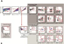
Fig.3.A

-
FC/FACS
-
Homo sapiens (Human)
Collected and cropped from PLoS One by CiteAb, provided under a CC-BY license
Image 1 of 2
In PLoS One on 23 August 2019 by Cavrois, M., Hilton, J. F., et al.
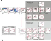
Fig.1.A

-
FC/FACS
-
Homo sapiens (Human)
Collected and cropped from PLoS One by CiteAb, provided under a CC-BY license
Image 1 of 2