Whole-exome sequencing of two unrelated kindreds with systemic autoimmune disease featuring antinuclear antibodies with IgG4 elevation uncovered an identical ultrarare heterozygous TNIP1Q333P variant segregating with disease. Mice with the orthologous Q346P variant developed antinuclear autoantibodies, salivary gland inflammation, elevated IgG2c, spontaneous germinal centers and expansion of age-associated B cells, plasma cells and follicular and extrafollicular helper T cells. B cell phenotypes were cell-autonomous and rescued by ablation of Toll-like receptor 7 (TLR7) or MyD88. The variant increased interferon-β without altering nuclear factor kappa-light-chain-enhancer of activated B cells signaling, and impaired MyD88 and IRAK1 recruitment to autophagosomes. Additionally, the Q333P variant impaired TNIP1 localization to damaged mitochondria and mitophagosome formation. Damaged mitochondria were abundant in the salivary epithelial cells of Tnip1Q346P mice. These findings suggest that TNIP1-mediated autoimmunity may be a consequence of increased TLR7 signaling due to impaired recruitment of downstream signaling molecules and damaged mitochondria to autophagosomes and may thus respond to TLR7-targeted therapeutics.
© 2024. The Author(s).
Product Citations: 14
A TNIP1-driven systemic autoimmune disorder with elevated IgG4.
In Nature Immunology on 1 September 2024 by Medhavy, A., Athanasopoulos, V., et al.
-
FC/FACS
-
Mus musculus (House mouse)
-
Immunology and Microbiology
Bank1 modulates the differentiation and molecular profile of key B cell populations in autoimmunity.
In JCI Insight on 20 August 2024 by Gómez Hernández, G., Domínguez, T., et al.
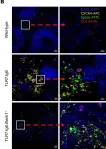
This study aimed at defining the role of the B cell adaptor protein BANK1 in the appearance of age-associated B cells (ABCs) in 2 SLE mouse models (TLR7.tg6 and imiquimod-induced mice), crossed with Bank1-/- mice. The absence of Bank1 led to a significant reduction in ABC levels, also affecting other B cell populations. To gain deeper insights into their differentiation pathway and the effect of Bank1 on B cell populations, a single-cell transcriptome assay was performed. In the TLR7.tg6 model, we identified 10 clusters within B cells, including an ABC-specific cluster that was decreased in Bank1-deficient mice. In its absence, ABCs exhibited an antiinflammatory gene expression profile, while being proinflammatory in Bank1-sufficient lupus-prone mice. Trajectory analyses revealed that ABCs originated from marginal zone and memory-like B cells, ultimately acquiring transcriptional characteristics associated with atypical memory cells and long-lived plasma cells. Also, Bank1 deficiency normalized the presence of naive B cells, which were nearly absent in lupus-prone mice. Interestingly, Bank1 deficiency significantly reduced a distinct cluster containing IFN-responsive genes. These findings underscore the critical role of Bank1 in ABC development, affecting early B cell stages toward ABC differentiation, and the presence of IFN-stimulated gene-containing B cells, both populations determinant for autoimmunity.
-
IHC-IF
-
Mus musculus (House mouse)
-
Immunology and Microbiology
In Frontiers in Cellular and Infection Microbiology on 17 July 2023 by Hernández-Cázares, F., Maqueda-Alfaro, R. A., et al.
Patients with Human Hyper IgM syndromes (HIGM) developed pulmonary and gastrointestinal infections since infancy and most patients have mutations in the CD40 ligand (CD40L) gene. Most HIGM patients compared to healthy subjects have higher/similar IgM and lower IgG, and IgA serum concentrations but gut antibody concentrations are unknown. CD40L on activated T-cells interacts with CD40 on B-cells, essential for the formation of germinal centres (GCs) inside secondary lymphoid organs (SLOs), where high-affinity antibodies, long-lived antibody-secreting plasma cells, and memory B-cells, are produced. C57BL6-CD40 ligand deficient mice (C57BL6-cd40l -/-), are a model of HIGM, because serum immunoglobulin concentrations parallel levels observed in HIGM patients and have higher faecal IgA concentrations. In mice, TGFβ and other cytokines induce IgA production.
To compare and evaluate B-cell populations and IgA-producing plasma cells in peritoneal lavage, non-gut-associated SLOs, spleen/inguinal lymph nodes (ILN), and gut-associated SLOs, mesenteric lymph nodes (MLN)/Peyer´s patches (PP) of unimmunised C57BL6-cd40l -/- and C57BL6-wild-type (WT) mice.
Peritoneal lavages, spleens, ILN, MLN, and PP from 8-10 weeks old C57BL6-cd40l -/- and WT mice, were obtained. Organ cryosections were analysed by immunofluorescence and B-cell populations and IgA-positive plasma cell suspensions by flow cytometry.
In unimmunised WT mice, GCs were only observed in the gut-associated SLOs, but GCs were absent in all C57BL6-cd40l -/- SLOs. PP and MLN of C57BL6-cd40l -/- mice exhibited a significantly higher number of IgA-producing cells than WT mice. In the spleen and ILN of C57BL6-cd40l- /- mice IgA-producing cells significantly decreased, while IgM-positive plasma cells increased. C57BL6-cd40l -/- B-1 cells were more abundant in all analysed SLOs, whereas in WT mice most B-1 cells were contained within the peritoneal cavity. C57BL6-cd40l -/- B-cells in MLN expressed a higher TGFβ receptor-1 than WT mice. Mouse strains small intestine microvilli (MV), have a similar frequency of IgA-positive cells.
Together our results confirm the role of PP and MLN as gut inductive sites, whose characteristic features are to initiate an IgA preferential immune response production in these anatomical sites even in the absence of GCs. IgA antibodies play a pivotal role in neutralising, eliminating, and regulating potential pathogens and microorganisms in the gut.
Copyright © 2023 Hernandez-Cazares, Maqueda-Alfaro, Lopez-Saucedo, Martinez-Barnetche, Yam-Puc, Estrada-Parra, Flores-Romo and Estrada-Garcia.
-
Immunology and Microbiology
In Gut Microbes on 15 June 2023 by Bessho, S., Grando, K. C. M., et al.
The Salmonella biofilm-associated amyloid protein, curli, is a dominant instigator of systemic inflammation and autoimmune responses following Salmonella infection. Systemic curli injections or infection of mice with Salmonella Typhimurium induce the major features of reactive arthritis, an autoimmune disorder associated with Salmonella infection in humans. In this study, we investigated the link between inflammation and microbiota in exacerbating autoimmunity. We studied C57BL/6 mice from two sources, Taconic Farms and Jackson Labs. Mice from Taconic Farms have been reported to have higher basal levels of the inflammatory cytokine IL - 17 than do mice from Jackson Labs due to the differences in their microbiota. When we systemically injected mice with purified curli, we observed a significant increase in diversity in the microbiota of Jackson Labs mice but not in that of the Taconic mice. In Jackson Labs, mice, the most striking effect was the expansion of Prevotellaceae. Furthermore, there were increases in the relative abundance of the family Akkermansiaceae and decreases in families Clostridiaceae and Muribaculaceae in Jackson Labs mice. Curli treatment led to significantly aggravated immune responses in the Taconic mice compared to Jackson Labs counterparts. Expression and production of IL - 1β, a cytokine known to promote IL - 17 production, as well as expression of Tnfa increased in the gut mucosa of Taconic mice in the first 24 hours after curli injections, which correlated with significant increases in the number of neutrophils and macrophages in the mesenteric lymph nodes. A significant increase in the expression of Ccl3 in colon and cecum of Taconic mice injected with curli was detected. Taconic mice injected with curli also had elevated levels of inflammation in their knees. Overall, our data suggest that autoimmune responses to bacterial ligands, such as curli, are amplified in individuals with a microbiome that promote inflammation.
-
FC/FACS
-
Mus musculus (House mouse)
-
Immunology and Microbiology
In JCI Insight on 22 February 2023 by Yick, L. W., Ma, O. K., et al.
Neuromyelitis optica spectrum disorders (NMOSD) are inflammatory autoimmune disorders of the CNS. IgG autoantibodies targeting the aquaporin-4 water channel (AQP4-IgGs) are the pathogenic effector of NMOSD. Dysregulated T follicular helper (Tfh) cells have been implicated in loss of B cell tolerance in autoimmune diseases. The contribution of Tfh cells to disease activity and therapeutic potential of targeting these cells in NMOSD remain unclear. Here, we established an autoimmune model of NMOSD by immunizing mice against AQP4 via in vivo electroporation. After AQP4 immunization, mice displayed AQP4 autoantibodies in blood circulation, blood-brain barrier disruption, and IgG infiltration in spinal cord parenchyma. Moreover, AQP4 immunization induced motor impairments and NMOSD-like pathologies, including astrocytopathy, demyelination, axonal loss, and microglia activation. These were associated with increased splenic Tfh, Th1, and Th17 cells; memory B cells; and plasma cells. Aqp4-deficient mice did not display motor impairments and NMOSD-like pathologies after AQP4 immunization. Importantly, abrogating ICOS/ICOS-L signaling using anti-ICOS-L antibody depleted Tfh cells and suppressed the response of Th1 and Th17 cells, memory B cells, and plasma cells in AQP4-immunized mice. These findings were associated with ameliorated motor impairments and spinal cord pathologies. This study suggests a role of Tfh cells in the pathophysiology of NMOSD in a mouse model with AQP4 autoimmunity and provides an animal model for investigating the immunological mechanisms underlying AQP4 autoimmunity and developing therapeutic interventions targeting autoimmune reactions in NMOSD.
-
Mus musculus (House mouse)
In JCI Insight on 20 August 2024 by Gómez Hernández, G., Domínguez, T., et al.
Fig.7.B

-
IHC-IF
-
Mus musculus (House mouse)
Collected and cropped from JCI Insight by CiteAb, provided under a CC-BY license
Image 1 of 1