Liver regeneration is supported by hepatocytes and, in certain conditions, biliary epithelial cells (BECs). BECs are facultative liver stem cells that form organoids in culture and engraft in damaged livers. However, BEC heterogeneity in the homeostatic liver remains to be fully elucidated. Here, we exploit systemic lentiviral vector (LV) administration to achieve efficient and lifelong gene transfer to BECs in mice. We find that LV-marked BECs retain organoid formation potential and predominantly respond to liver damage; however, they are less clonogenic and display a hepatocyte-primed transcriptome compared to untransduced BECs. We thus identify a BEC subset committed to hepatocyte lineage in the absence of liver damage, characterized by a transcriptional network orchestrated by hepatocyte nuclear factor 4α. We also report in vivo targeting of such BECs in non-human primates. This work highlights intrinsic BEC heterogeneity and that in vivo LV gene transfer to the liver may persist following BEC-mediated repair of hepatic damage.
Copyright © 2025 The Author(s). Published by Elsevier Inc. All rights reserved.
Product Citations: 27
In Cell Reports on 25 March 2025 by Milani, M., Starinieri, F., et al.
In Stem Cell Research & Therapy on 1 March 2025 by Yao, Y., Luo, Y., et al.
Stem cells play a pivotal role in tissue regeneration and repair. Skeletal muscle comprises two main stem cells: muscle stem cells (MuSCs) and fibro-adipogenic progenitors (FAPs). FAPs are essential for maintaining the regenerative milieu of muscle tissue and modulating the activation of muscle satellite cells. However, during acute skeletal muscle injury, the alterations and mechanisms of action of FAPs remain unclear.
we employed the GEO database for bioinformatics analysis of skeletal muscle injury. A skeletal muscle injury model was established through cardiotoxin (CTX, 10µM, 50µL) injection into the tibialis anterior (TA) of C57BL/6 mice. Three days post-injury, we extracted the TA, isolated FAPs (CD31-CD45-PDGFRα+Sca-1+), and assessed the senescence phenotype through SA-β-Gal staining and Western blot. Additionally, we established a co-culture system to evaluate the capacity of FAPs to facilitate MuSCs differentiation. Finally, we alleviated the senescent of FAPs through in vitro (100 µM melatonin, 5 days) and in vivo (20 mg/kg/day melatonin, 15 days) administration experiments, confirming melatonin's pivotal role in the regeneration and repair processes of skeletal muscle.
In single-cell RNA sequencing analysis, we discovered the upregulation of senescence-related pathways in FAPs following injury. Immunofluorescence staining revealed the co-localization of FAPs and senescent markers in injured muscles. We established the CTX injury model and observed a reduction in the number of FAPs post-injury, accompanied by the manifestation of a senescent phenotype. Melatonin treatment was found to attenuate the injury-induced senescence of FAPs. Further co-culture experiments revealed that melatonin facilitated the restoration of FAPs' capacity to promote myoblast differentiation. Through GO and KEGG analysis, we found that the administration of melatonin led to the upregulation of AMPK pathway in FAPs, a pathway associated with antioxidant stress response. Finally, drug administration experiments corroborated that melatonin enhances skeletal muscle regeneration and repair by alleviating FAP senescence in vivo.
In this study, we first found FAPs underwent senescence and redox homeostasis imbalance after injury. Next, we utilized melatonin to enhance FAPs regenerative and repair capabilities by activating AMPK signaling pathway. Taken together, this work provides a novel theoretical foundation for treating skeletal muscle injury.
© 2025. The Author(s).
-
Stem Cells and Developmental Biology
Endurance exercise remodels skeletal muscle by suppressing Ythdf1-mediated myostatin expression.
In Cell Death & Disease on 13 February 2025 by Huang, X., Xu, C., et al.
Exercise can improve health via skeletal muscle remodeling. Elucidating the underlying mechanism may lead to new therapeutics for aging-related loss of skeletal muscle mass. Here, we show that endurance exercise suppresses expression of YT521-B homology domain family (Ythdf1) in skeletal muscle, which recognizes the N6-methyladenosine (m6A). Ythdf1 deletion phenocopies endurance exercise-induced muscle hypertrophy in mice increases muscle mitochondria content and type I fiber specification. At the molecular level, Ythdf1 recognizes and promotes the translation of m6A-modified Mstn mRNA, which encodes a muscle growth inhibitor, Myostatin. Loss of Ythdf1 leads to hyperactivation of skeletal muscle stem cells (MuSCs), also called satellite cells (SCs), enhancing muscle growth and injury-induced regeneration. Our data reveal Ythdf1 as a key regulator of skeletal muscle homeostasis, provide insights into the mechanism by which endurance exercise promotes skeletal muscle remodeling and highlight potential strategies to prevent aging-related muscle degeneration.
© 2025. The Author(s).
-
Mus musculus (House mouse)
-
Cell Biology
Depleting myeloid-biased haematopoietic stem cells rejuvenates aged immunity.
In Nature on 1 April 2024 by Ross, J. B., Myers, L. M., et al.
Ageing of the immune system is characterized by decreased lymphopoiesis and adaptive immunity, and increased inflammation and myeloid pathologies1,2. Age-related changes in populations of self-renewing haematopoietic stem cells (HSCs) are thought to underlie these phenomena3. During youth, HSCs with balanced output of lymphoid and myeloid cells (bal-HSCs) predominate over HSCs with myeloid-biased output (my-HSCs), thereby promoting the lymphopoiesis required for initiating adaptive immune responses, while limiting the production of myeloid cells, which can be pro-inflammatory4. Ageing is associated with increased proportions of my-HSCs, resulting in decreased lymphopoiesis and increased myelopoiesis3,5,6. Transfer of bal-HSCs results in abundant lymphoid and myeloid cells, a stable phenotype that is retained after secondary transfer; my-HSCs also retain their patterns of production after secondary transfer5. The origin and potential interconversion of these two subsets is still unclear. If they are separate subsets postnatally, it might be possible to reverse the ageing phenotype by eliminating my-HSCs in aged mice. Here we demonstrate that antibody-mediated depletion of my-HSCs in aged mice restores characteristic features of a more youthful immune system, including increasing common lymphocyte progenitors, naive T cells and B cells, while decreasing age-related markers of immune decline. Depletion of my-HSCs in aged mice improves primary and secondary adaptive immune responses to viral infection. These findings may have relevance to the understanding and intervention of diseases exacerbated or caused by dominance of the haematopoietic system by my-HSCs.
© 2024. The Author(s), under exclusive licence to Springer Nature Limited.
-
Immunology and Microbiology
-
Stem Cells and Developmental Biology
In Journal of Neuroimmunology on 15 March 2024 by Trevino, T. N., Almousawi, A. A., et al.
Blood-brain barrier (BBB) permeability can cause neuroinflammation and cognitive impairment. Caveolin-1 (Cav-1) critically regulates BBB permeability, but its influence on the BBB and consequent neurological outcomes in respiratory viral infections is unknown. We used Cav-1-deficient mice with genetically encoded fluorescent endothelial tight junctions to determine how Cav-1 influences BBB permeability, neuroinflammation, and cognitive impairment following respiratory infection with mouse adapted (MA10) SARS-CoV-2 as a model for COVID-19. We found that SARS-CoV-2 infection increased brain endothelial Cav-1 and increased transcellular BBB permeability to albumin, decreased paracellular BBB Claudin-5 tight junctions, and caused T lymphocyte infiltration in the hippocampus, a region important for learning and memory. Concordantly, we observed learning and memory deficits in SARS-CoV-2 infected mice. Importantly, genetic deficiency in Cav-1 attenuated transcellular BBB permeability and paracellular BBB tight junction losses, T lymphocyte infiltration, and gliosis induced by SARS-CoV-2 infection. Moreover, Cav-1 KO mice were protected from the learning and memory deficits caused by SARS-CoV-2 infection. These results establish the contribution of Cav-1 to BBB permeability and behavioral dysfunction induced by SARS-CoV-2 neuroinflammation.
Copyright © 2024 The Authors. Published by Elsevier B.V. All rights reserved.
-
Cardiovascular biology
-
COVID-19
-
Immunology and Microbiology
-
Neuroscience
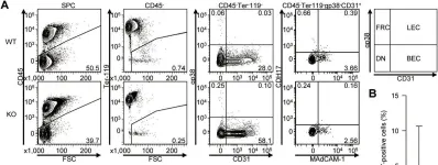
In PLoS One on 23 January 2015 by Funakoshi, S., Shimizu, T., et al.
Fig.6.A

-
FC/FACS
-
Collected and cropped from PLoS One by CiteAb, provided under a CC-BY license
Image 1 of 1