Severe acute respiratory syndrome coronavirus 2 (SARS-CoV-2) RNA generally becomes undetectable in upper airways after a few days or weeks postinfection. Here we used a model of viral infection in macaques to address whether SARS-CoV-2 persists in the body and which mechanisms regulate its persistence. Replication-competent virus was detected in bronchioalveolar lavage (BAL) macrophages beyond 6 months postinfection. Viral propagation in BAL macrophages occurred from cell to cell and was inhibited by interferon-γ (IFN-γ). IFN-γ production was strongest in BAL NKG2r+CD8+ T cells and NKG2Alo natural killer (NK) cells and was further increased in NKG2Alo NK cells after spike protein stimulation. However, IFN-γ production was impaired in NK cells from macaques with persisting virus. Moreover, IFN-γ also enhanced the expression of major histocompatibility complex (MHC)-E on BAL macrophages, possibly inhibiting NK cell-mediated killing. Macaques with less persisting virus mounted adaptive NK cells that escaped the MHC-E-dependent inhibition. Our findings reveal an interplay between NK cells and macrophages that regulated SARS-CoV-2 persistence in macrophages and was mediated by IFN-γ.
© 2023. The Author(s).
Product Citations: 10
SARS-CoV-2 viral persistence in lung alveolar macrophages is controlled by IFN-γ and NK cells.
In Nature Immunology on 1 December 2023 by Huot, N., Planchais, C., et al.
-
COVID-19
-
Immunology and Microbiology
Optimized Nonviral Gene Disruption in Primary Murine and Human Myeloid Cells.
In Methods in Molecular Biology (Clifton, N.J.) on 12 March 2023 by Freund, E. C., Haag, S. M., et al.
Genetically engineered myeloid cells such as monocytes, macrophages, and dendritic cells have broad applications in basic and translational research. Their central roles in innate and adaptive immunity make them attractive as putative therapeutic cell products. However, efficient gene editing of primary myeloid cells presents unique challenges owing to their sensitivity to foreign nucleic acids and poor editing efficiencies using current methodologies (Hornung et al., Science 314:994-997, 2006; Coch et al., PLoS One 8:e71057, 2013; Bartok and Hartmann, Immunity 53:54-77, 2020; Hartmann, Adv Immunol 133:121-169, 2017; Bobadilla et al., Gene Ther 20:514-520, 2013; Schlee and Hartmann, Nat Rev Immunol 16:566-580, 2016; Leyva et al., BMC Biotechnol 11:13, 2011). This chapter describes nonviral CRISPR-mediated gene knockout in primary human and murine monocytes as well as monocyte-derived or bone marrow-derived macrophages and dendritic cells. Electroporation-mediated delivery of recombinant Cas9 complexed with synthetic guide RNAs can be applied for population-level disruption of single or multiple gene targets.
© 2023. The Author(s), under exclusive license to Springer Science+Business Media, LLC, part of Springer Nature.
-
Biochemistry and Molecular biology
Amelioration of Lupus Serum-Induced Skin Inflammation in CD64-Deficient Mice.
In Frontiers in Immunology on 12 March 2022 by Jiang, L., Han, X., et al.
Systemic lupus erythematosus (SLE) is a heterogeneous autoimmune disorder characterized by high autoantibodies levels and multiorgan tissue damage. The current study investigated the role of CD64 in SLE patients and animal models. According to a flow cytometry study, SLE patients showed an increase in CD64 expression in circulating monocytes. There was a correlation between CD64 and SLEDAI, blood urea nitrogen levels, and anti-Sm antibodies. In skin lesions of lupus MRL/lpr mice, there was high IgG deposition and CD64 expression. In vitro, cytokines IL-10 and IFN-γ upregulated CD64 expression in monocytes/macrophages that was inhibited by glucocorticoids. In CD64-deficient mice, skin inflammation induced by lupus serum was reduced. Furthermore, activation of spleen tyrosine kinase (Syk), Akt, and extracellular signal-regulated kinase (Erk) was inhibited in CD64-deficient monocytes. The results suggest that CD64 could be a biomarker for observing SLE progression, as well as a mechanistic checkpoint in lupus pathogenesis.
Copyright © 2022 Jiang, Han, Qiu, Yu, Feng, Wang, Duan and Deng.
-
Immunology and Microbiology
ACE2 can act as the secondary receptor in the FcγR-dependent ADE of SARS-CoV-2 infection.
In IScience on 21 January 2022 by Wang, Z., Deng, T., et al.
It is unknown whether antibody-mediated enhancement (ADE) contributes to the pathogenesis of COVID-19, and the conditions for ADE needs to be elucidated. We demonstrated that without inducing an ACE2-independent ADE on Raji cells, the neutralizing antibody CB6, a mouse anti-S1 serum and convalescent plasma, induced ADE on cells expressing FcγRIIA/CD32A and low levels of endogenous ACE2. ADE occurred at sub-neutralizing antibody concentrations, indicating that unneutralized S protein was required for ADE. The enhanced infectivity of 614G variant was higher than that of 614D wildtype in the presence of antibodies, further suggesting that ADE may be influenced by virus strains with different ACE2-binding affinity. Finally, knockdown of ACE2 or treatment with a fusion-inhibition peptide EK1C4 significantly reduced ADE. In conclusion, we identified an ADE mechanism mediated by neutralizing antibodies against SARS-CoV-2. ACE2 may act as a secondary receptor required for the antibody- and FcγR-mediated enhanced entry of SARS-CoV-2.
© 2021 The Author(s).
-
COVID-19
-
Immunology and Microbiology
In Viruses on 11 December 2021 by Shen, X. R., Li, Q., et al.
Patients with COVID-19 generally raise antibodies against SARS-CoV-2 following infection, and the antibody level is positively correlated to the severity of disease. Whether the viral antibodies exacerbate COVID-19 through antibody-dependent enhancement (ADE) is still not fully understood. Here, we conducted in vitro assessment of whether convalescent serum enhanced SARS-CoV-2 infection or induced excessive immune responses in immune cells. Our data revealed that SARS-CoV-2 infection of primary B cells, macrophages and monocytes, which express variable levels of FcγR, could be enhanced by convalescent serum from COVID-19 patients. We also determined the factors associated with ADE, and found which showed a time-dependent but not viral-dose dependent manner. Furthermore, the ADE effect is not associated with the neutralizing titer or RBD antibody level when testing serum samples collected from different patients. However, it is higher in a medium level than low or high dilutions in a given sample that showed ADE effect, which is similar to dengue. Finally, we demonstrated more viral genes or dysregulated host immune gene expression under ADE conditions compared to the no-serum infection group. Collectively, our study provides insight into the understanding of an association of high viral antibody titer and severe lung pathology in severe patients with COVID-19.
-
FC/FACS
-
COVID-19
-
Immunology and Microbiology
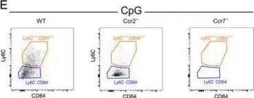
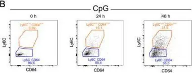
In Sci Rep on 20 July 2017 by De Koker, S., Van Hoecke, L., et al.
Fig.6.E

-
FC/FACS
-
Collected and cropped from Sci Rep by CiteAb, provided under a CC-BY license
Image 1 of 2
In Sci Rep on 20 July 2017 by De Koker, S., Van Hoecke, L., et al.
Fig.6.B

-
FC/FACS
-
Collected and cropped from Sci Rep by CiteAb, provided under a CC-BY license
Image 1 of 2