Dental pulp stem cells (DPSCs) have demonstrated remarkable potential in enhancing peripheral nerve regeneration, though the precise mechanisms remain largely unknown. This study investigates how DPSCs alleviate Schwann cell pyroptosis and restore mitochondrial homeostasis through intercellular mitochondrial transfer. In a crab-eating macaque model, we first observed that DPSC-loaded nerve conduits significantly promoted long-term nerve regeneration, facilitating tissue proliferation and myelin recovery. We further established a rat facial nerve injury (FNI) model and found that DPSC treatment reduced pyroptosis and mitochondrial ROS production in Schwann cells. A pivotal mitochondrial protective mechanism, resembling the effects of a ROS-targeted inhibitor, involved the transfer of mitochondria from DPSCs to pyroptosis-induced Schwann cells via tunneling nanotubes, while blocking intercellular junctions or mitochondrial function diminished the therapeutic effects. TNFα secreted by pyroptosis-induced Schwann cells activated the NF-κB pathway in DPSCs, enhancing mitochondrial transfer and adaptive stress responses, thereby promoting mitochondrial protection against pyroptosis in Schwann cells, as reflected in the improved therapeutic efficacy of TNFα-preconditioned DPSCs in the FNI model. These findings unveil a mechanism through which DPSCs foster nerve regeneration via mitochondrial transfer, presenting a promising strategy for enhancing stem cell-based therapies for nerve injuries.
© 2025 The Authors.
Product Citations: 91
In Bioactive Materials on 1 May 2025 by Zheng, X., Wang, J., et al.
-
FC/FACS
-
Cell Biology
-
Neuroscience
-
Stem Cells and Developmental Biology
In Scientific Reports on 24 February 2025 by Bulut, O., Genç, D., et al.
The aim of this study was to investigate the regenerative effect of lyophilized dental follicle mesenchymal stem cells (DF-MSCs) combined with rat platelet-rich fibrin (PRF) on geriatric skin wounds. Human DF-MSCs which were isolated from the wisdom teeth of healthy donors and PRF were mixed and incubated in a 37 °C incubator for 1-2 h containing 1 million cells in 150 mg PRF. The mixture was suspended in a freeze-drying solution and then lyophilized. Wounds were created on the back skin of Wistar albino rats using a 6 mm punch. Lyophilized DF-MSCs, PRF, or PRF + DF-MSCs were applied to the wounds of rats. On the 15th day, the wound area was histopathologically evaluated in rats. Blood samples from rats were analyzed for total antioxidant status (TAOS), and inflammatory cytokine levels using ELISA. In both young and geriatric rats treated with lyophilized PRF + DF-MSCs, wound area began to significantly decrease from the 10th day compared to the untreated group (p < 0.05). Histopathological examination revealed that in the lyophilized PRF + DF-MSCs treated groups, epithelial integrity and scarless healing significantly increased compared to the untreated groups (p < 0.05). There were no significant differences in TAOS, total oxidant status (TOS), tumor necrosis factor (TNF), interleukin-6 (IL6), and hydroxyproline levels in serum samples from young rats on the 15th day. In geriatric rats, hydroxyproline (HYPS) levels were increased in the DF-MSC and PRF + DF-MSC groups (p < 0.01), TNF was significantly elevated in PRF geriatric group and IL6 was increased in the PRF group compared to the control group (p = 0.01). Lyophilized PRF + DF-MSCs, which is a shelf-stable and ready-to-use product, hold promise, especially for traumatic wounds in geriatric individuals with longer healing times.
© 2025. The Author(s).
-
Stem Cells and Developmental Biology
Identification of human cranio-maxillofacial skeletal stem cells for mandibular development.
In Science Advances on 3 January 2025 by Wang, Z., Wang, K., et al.
Compared with long bone that arises from the mesoderm, the major portion of the maxillofacial bones and the front bone of the skull are derived from cranial neural crest cells and undergo intramembranous ossification. Human skeletal stem cells have been identified in embryonic and fetal long bones. Here, we describe a single-cell atlas of the human embryonic mandible and identify a population of cranio-maxillofacial skeletal stem cells (CMSSCs). These CMSSCs are marked by interferon-induced transmembrane protein 5 (IFITM5) and are specifically located around the periosteum of the jawbone and frontal bone. Additionally, these CMSSCs exhibit strong self-renewal and osteogenic differentiation capacities but lower chondrogenic differentiation potency, mediating intramembranous bone formation without cartilage formation. IFITM5+ cells are also observed in the adult jawbone and exhibit functions similar to those of embryonic CMSSCs. Thus, this study identifies CMSSCs that orchestrate the intramembranous ossification of cranio-maxillofacial bones, providing a deeper understanding of cranio-maxillofacial skeletal development and promising seed cells for bone repair.
-
Stem Cells and Developmental Biology
IL-10RA governor the expression of IDO in the instruction of lymphocyte immunity.
In British Journal of Cancer on 1 January 2025 by Tai, T. S., Hsu, D. W., et al.
Indoleamine 2,3-dioxygenase (IDO) impairs anti-pathogen and anti-tumour immunity. Mesenchymal stem cells (MSCs) modulate immunity via IDO but also suppress IFN-γ. While MSC IDO induction by IFN-γ is established, other drivers in this immunosuppressive setting remain unknown.
Human bone marrow mesenchymal stem cells (MSCs) with IDO or IL-10RA knockdown were co-cultured with healthy donor T cells to assess immunosuppression. PDAC organoid anticancer activity was also tested in these co-cultures.
Co-culturing MSCs with T cells in an IL-10RA-enriched environment enhances IDO expression, resulting in T cell suppression. Moreover, IL-10RA-positive MSCs collected from co-cultures with IL-10 supplementation show increased IDO expression. Conversely, MSCs with IL-10RA knockdown exhibit a significant reduction in IDO RNA and protein expression, as well as STAT3 phosphorylation status, which is a known upstream signalling pathway in IDO gene regulation, in T cell co-cultures. Down-regulation of IL-10RA also inhibits IDO activity in MSCs, resulting in reduced T cell suppression, and enabling the co-cultured T cells to kill PDAC organoids.
Our research reveals IL-10RA as a pharmacological target in stromal cells for enhancing T cell-mediated PDAC eradication by downregulating IDO via blocked IL-10/IL-10RA signalling in MSCs. This advances IL-10RA interference in the tumour microenvironment (TME) to restore T cell cytotoxicity against cancers.
© 2024. The Author(s).
-
FC/FACS
-
Homo sapiens (Human)
-
Cancer Research
-
Immunology and Microbiology
Development and analysis of scaffold-free adipose spheroids.
In Adipocyte on 1 December 2024 by Liszewski, J. N., Klingelhutz, A. J., et al.
Adipose tissue plays a crucial role in metabolic syndrome, autoimmune diseases, and many cancers. Because of adipose's role in so many aspects of human health, there is a critical need for in vitro models that replicate adipose architecture and function. Traditional monolayer models, despite their convenience, are limited, showing heterogeneity and functional differences compared to 3D models. While monolayer cultures struggle with detachment and inefficient differentiation, healthy adipocytes in 3D culture accumulate large lipid droplets, secrete adiponectin, and produce low levels of inflammatory cytokines. The shift from monolayer models to more complex 3D models aims to better replicate the physiology of healthy adipose tissue in culture. This study introduces a simple and accessible protocol for generating adipose organoids using a scaffold-free spheroid model. The method, utilizing either 96-well spheroid plates or agarose micromolds, demonstrates increased throughput, uniformity, and ease of handling compared to previous techniques. This protocol allows for diverse applications, including drug testing, toxin screening, tissue engineering, and co-culturing. The choice between the two methods depends on the experimental goals, with the 96-well plate providing individualized control and the micromold offering scale advantages. The outlined protocol covers isolation, expansion, and characterization of stromal vascular fraction cells, followed by detailed steps for spheroid formation and optional downstream analyses.
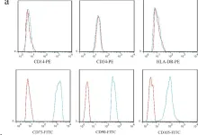
In Stem Cell Res Ther on 5 February 2022 by Ye, F., Li, J., et al.
Fig.1.A

-
FC/FACS
-
Collected and cropped from Stem Cell Res Ther by CiteAb, provided under a CC-BY license
Image 1 of 1