Peripheral nerve injuries can lead to lasting functional impairments, impacting movement and quality of life. FK-506, a widely used immunosuppressant, has demonstrated potential in promoting nerve regeneration in addition to its immunosuppressive effects. This study investigates the use of a local reservoir flap to deliver FK-506 directly to the nerve injury site, aiming to enhance nerve regeneration while minimizing systemic immunosuppression.
Sciatic nerve injuries were surgically induced in 24 rats, which were divided into control, 0.5 mg/kg FK-506 (Exp 1), and 2.0 mg/kg FK-506 (Exp 2) groups. A superficial inferior epigastric artery flap served as a reservoir for FK-506, allowing direct delivery to the injury site. FK-506 was administered intermittently over a 4-week period. Outcomes included the Sciatic Functional Index (SFI), muscle recovery (width and weight), nerve morphology, expression of neurogenic markers such as GDNF, immune cell counts, and body weight.
Exp 1 (0.5 mg/kg) demonstrated significant improvements in SFI, GDNF expression, and muscle width compared to the control and high-dose groups. These findings suggest that FK-506 administration via a reservoir flap, particularly at a lower dose, supports effective nerve regeneration. Additionally, FK-506 treatment did not result in significant changes in immune cell profiles or body weight, indicating minimal systemic effects.
Localized FK-506 administration via a reservoir flap effectively enhances peripheral nerve regeneration and minimizes systemic immunosuppression, making it a promising approach for clinical application in treating peripheral nerve injuries.
© Copyright: Yonsei University College of Medicine 2024.
Product Citations: 6
In Yonsei Medical Journal on 1 December 2024 by Hong, J. W., Lim, J. H., et al.
-
Rattus norvegicus (Rat)
-
Neuroscience
Functional differences in airway dendritic cells determine susceptibility to IgE-sensitization.
In Immunology and Cell Biology on 1 March 2018 by Leffler, J., Mincham, K. T., et al.
Respiratory IgE-sensitization to innocuous antigens increases the risk for developing diseases such as allergic asthma. Dendritic cells (DC) residing in the airways orchestrate the immune response following antigen exposure and their ability to sample and present antigens to naïve T cells in airway draining lymph nodes contributes to allergen-specific IgE-sensitization. In order to characterize inhaled antigen capture and presentation by DC subtypes in vivo, we used an adjuvant-free respiratory sensitization model using two genetically distinct rat strains, one of which is naturally resistant and the other naturally susceptible to allergic sensitization. Upon multiple exposures to ovalbumin (OVA), the susceptible strain developed OVA-specific IgE and airway inflammation, whereas the resistant strain did not. Using fluorescently tagged OVA and flow cytometry, we demonstrated significant differences in antigen uptake efficiency and presentation associated with either IgE-sensitization or resistance to allergen exposures in respective strains. We further identified CD4+ conventional DC (cDC) as the subset involved in airway antigen sampling in both strains, however, CD4+ cDC in the susceptible strain were less efficient in OVA sampling and displayed increased MHC-II expression compared with the resistant strain. This was associated with generation of an exaggerated Th2 response and a deficiency of airway regulatory T cells in the susceptible strain. These data suggest that subsets of cDC are able to induce either sensitization or resistance to inhaled antigens as determined by genetic background, which may provide an underlying basis for genetically determined susceptibility to respiratory allergic sensitization and IgE production in susceptible individuals.
© 2017 Australasian Society for Immunology Inc.
-
Immunology and Microbiology
Neuregulin-1 elicits a regulatory immune response following traumatic spinal cord injury.
In Journal of Neuroinflammation on 21 February 2018 by Alizadeh, A., Santhosh, K. T., et al.
Spinal cord injury (SCI) triggers a robust neuroinflammatory response that governs secondary injury mechanisms with both degenerative and pro-regenerative effects. Identifying new immunomodulatory therapies to promote the supportive aspect of immune response is critically needed for the treatment of SCI. We previously demonstrated that SCI results in acute and permanent depletion of the neuronally derived Neuregulin-1 (Nrg-1) in the spinal cord. Increasing the dysregulated level of Nrg-1 through acute intrathecal Nrg-1 treatment enhanced endogenous cell replacement and promoted white matter preservation and functional recovery in rat SCI. Moreover, we identified a neuroprotective role for Nrg-1 in moderating the activity of resident astrocytes and microglia following injury. To date, the impact of Nrg-1 on immune response in SCI has not yet been investigated. In this study, we elucidated the effect of systemic Nrg-1 therapy on the recruitment and function of macrophages, T cells, and B cells, three major leukocyte populations involved in neuroinflammatory processes following SCI.
We utilized a clinically relevant model of moderately severe compressive SCI in female Sprague-Dawley rats. Nrg-1 (2 μg/day) or saline was delivered subcutaneously through osmotic mini-pumps starting 30 min after SCI. We conducted flow cytometry, quantitative real-time PCR, and immunohistochemistry at acute, subacute, and chronic stages of SCI to investigate the effects of Nrg-1 treatment on systemic and spinal cord immune response as well as cytokine, chemokine, and antibody production.
We provide novel evidence that Nrg-1 promotes a pro-regenerative immune response after SCI. Bioavailability of Nrg-1 stimulated a regulatory phenotype in T and B cells and augmented the population of M2 macrophages in the spinal cord and blood during the acute and chronic stages of SCI. Importantly, Nrg-1 fostered a more balanced microenvironment in the injured spinal cord by attenuating antibody deposition and expression of pro-inflammatory cytokines and chemokines while upregulating pro-regenerative mediators.
We provide the first evidence of a significant regulatory role for Nrg-1 in neuroinflammation after SCI. Importantly, the present study establishes the promise of systemic Nrg-1 treatment as a candidate immunotherapy for traumatic SCI and other CNS neuroinflammatory conditions.
-
Rattus norvegicus (Rat)
-
Immunology and Microbiology
-
Neuroscience
In Journal of Leukocyte Biology on 1 April 2017 by Quek, H., Luff, J., et al.
Mutations in the ataxia-telangiectasia (A-T)-mutated (ATM) gene give rise to the human genetic disorder A-T, characterized by immunodeficiency, cancer predisposition, and neurodegeneration. Whereas a series of animal models recapitulate much of the A-T phenotype, they fail to present with ataxia or neurodegeneration. We describe here the generation of an Atm missense mutant [amino acid change of leucine (L) to proline (P) at position 2262 (L2262P)] rat by intracytoplasmic injection (ICSI) of mutant sperm into oocytes. Atm-mutant rats (AtmL2262P/L2262P ) expressed low levels of ATM protein, suggesting a destabilizing effect of the mutation, and had a significantly reduced lifespan compared with Atm+/+ Whereas these rats did not show cerebellar atrophy, they succumbed to hind-limb paralysis (45%), and the remainder developed tumors. Closer examination revealed the presence of both dsDNA and ssDNA in the cytoplasm of cells in the hippocampus, cerebellum, and spinal cord of AtmL2262P/L2262P rats. Significantly increased levels of IFN-β and IL-1β in all 3 tissues were indicative of DNA damage induction of the type 1 IFN response. This was further supported by NF-κB activation, as evidenced by p65 phosphorylation (P65) and translocation to the nucleus in the spinal cord and parahippocampus. Other evidence of neuroinflammation in the brain and spinal cord was the loss of motor neurons and the presence of increased activation of microglia. These data provide support for a proinflammatory phenotype that is manifested in the Atm mutant rat as hind-limb paralysis. This mutant represents a useful model to investigate the importance of neuroinflammation in A-T.
© Society for Leukocyte Biology.
-
Genetics
-
Immunology and Microbiology
-
Neuroscience
Functional characterization of human T cell hyporesponsiveness induced by CTLA4-Ig.
In PLoS ONE on 11 April 2015 by Rochman, Y., Yukawa, M., et al.
During activation, T cells integrate multiple signals from APCs and cytokine milieu. The blockade of these signals can have clinical benefits as exemplified by CTLA4-Ig, which blocks interaction of B7 co-stimulatory molecules on APCs with CD28 on T cells. Variants of CTLA4-Ig, abatacept and belatacept are FDA approved as immunosuppressive agents in arthritis and transplantation, yet murine studies suggested that CTLA4-Ig could be beneficial in a number of other diseases. However, detailed analysis of human CD4 cell hyporesponsivness induced by CTLA4-Ig has not been performed. Herein, we established a model to study the effect of CTLA4-Ig on the activation of human naïve T cells in a human mixed lymphocytes system. Comparison of human CD4 cells activated in the presence or absence of CTLA4-Ig showed that co-stimulation blockade during TCR activation does not affect NFAT signaling but results in decreased activation of NF-κB and AP-1 transcription factors followed by a profound decrease in proliferation and cytokine production. The resulting T cells become hyporesponsive to secondary activation and, although capable of receiving TCR signals, fail to proliferate or produce cytokines, demonstrating properties of anergic cells. However, unlike some models of T cell anergy, these cells did not possess increased levels of the TCR signaling inhibitor CBLB. Rather, the CTLA4-Ig-induced hyporesponsiveness was associated with an elevated level of p27kip1 cyclin-dependent kinase inhibitor.
-
Immunology and Microbiology
In PLoS One on 16 August 2014 by Abbondanzo, S. J. & Chang, S. L.
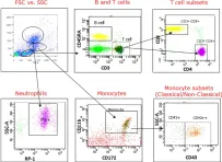
Fig.1.A

-
FC/FACS
-
Rattus norvegicus (Rat)
Collected and cropped from PLoS One by CiteAb, provided under a CC-BY license
Image 1 of 1
