Control of cell proliferation is critical for the lymphocyte life cycle. However, little is known about how stage-specific alterations in cell cycle behavior drive proliferation dynamics during T cell development. Here, we employed in vivo dual-nucleoside pulse labeling combined with the determination of DNA replication over time as well as fluorescent ubiquitination-based cell cycle indicator mice to establish a quantitative high-resolution map of cell cycle kinetics of thymocytes. We developed an agent-based mathematical model of T cell developmental dynamics. To generate the capacity for proliferative bursts, cell cycle acceleration followed a "stretch model" characterized by the simultaneous and proportional contraction of both G1 and S phases. Analysis of cell cycle phase dynamics during regeneration showed tailored adjustments of cell cycle phase dynamics. Taken together, our results highlight intrathymic cell cycle regulation as an adjustable system to maintain physiologic tissue homeostasis and foster our understanding of dysregulation of the T cell developmental program.
Copyright © 2024 The Author(s). Published by Elsevier Inc. All rights reserved.
Product Citations: 27
In Cell Reports on 28 January 2025 by Kunze-Schumacher, H., Verheyden, N. A., et al.
Leukocytes use endothelial membrane tunnels to extravasate the vasculature
Preprint on BioRxiv : the Preprint Server for Biology on 30 October 2024 by van der Meer, W. J., van Steen, A. C., et al.
ABSTRACT Upon inflammation, leukocytes extravasate through endothelial cells. When they extravasate in a paracellular manner, it is generally accepted that neighbouring endothelial cells physically disconnect to open cell-cell junctions, allowing leukocytes to cross. When carefully examining endothelial junctions, we found a partial membrane overlap of endothelial cells beyond VE-cadherin distribution. These overlaps are regulated by actin polymerization and, although marked by, do not require PECAM-1, nor VE-cadherin. Neutrophils prefer wider membrane overlaps as exit sites. Detailed 3D analysis of endothelial membrane dynamics during paracellular neutrophil transmigration in real-time, at high spatiotemporal resolution using resonant confocal and lattice light-sheet imaging, revealed that overlapping endothelial membranes form a tunnel during neutrophil transmigration. These tunnels are formed by the neutrophil lifting the membrane of the upper endothelial cell while indenting and crawling over the membrane of the underlying endothelial cell. Our work shows that endothelial cells do not simply retract upon passage of neutrophils but provide membrane tunnels, allowing neutrophils to extravasate. This discovery defines the 3D multicellular architecture in which the paracellular transmigration of neutrophils occurs.
C3aR-initiated signaling is a critical mechanism of podocyte injury in membranous nephropathy.
In JCI Insight on 16 January 2024 by Zhang, Q., Bin, S., et al.
The deposition of antipodocyte autoantibodies in the glomerular subepithelial space induces primary membranous nephropathy (MN), the leading cause of nephrotic syndrome worldwide. Taking advantage of the glomerulus-on-a-chip system, we modeled human primary MN induced by anti-PLA2R antibodies. Here we show that exposure of primary human podocytes expressing PLA2R to MN serum results in IgG deposition and complement activation on their surface, leading to loss of the chip permselectivity to albumin. C3a receptor (C3aR) antagonists as well as C3AR gene silencing in podocytes reduced oxidative stress induced by MN serum and prevented albumin leakage. In contrast, inhibition of the formation of the membrane-attack-complex (MAC), previously thought to play a major role in MN pathogenesis, did not affect permselectivity to albumin. In addition, treatment with a C3aR antagonist effectively prevented proteinuria in a mouse model of MN, substantiating the chip findings. In conclusion, using a combination of pathophysiologically relevant in vitro and in vivo models, we established that C3a/C3aR signaling plays a critical role in complement-mediated MN pathogenesis, indicating an alternative therapeutic target for MN.
In EMBO Reports on 9 January 2023 by Grönloh, M. L. B., Arts, J. J. G., et al.
Upon inflammation, leukocytes leave the circulation by crossing the endothelial monolayer at specific transmigration "hotspot" regions. Although these regions support leukocyte transmigration, their functionality is not clear. We found that endothelial hotspots function to limit vascular leakage during transmigration events. Using the photoconvertible probe mEos4b, we traced back and identified original endothelial transmigration hotspots. Using this method, we show that the heterogeneous distribution of ICAM-1 determines the location of the transmigration hotspot. Interestingly, the loss of ICAM-1 heterogeneity either by CRISPR/Cas9-induced knockout of ICAM-1 or equalizing the distribution of ICAM-1 in all endothelial cells results in the loss of TEM hotspots but not necessarily in reduced TEM events. Functionally, the loss of endothelial hotspots results in increased vascular leakage during TEM. Mechanistically, we demonstrate that the 3 extracellular Ig-like domains of ICAM-1 are crucial for hotspot recognition. However, the intracellular tail of ICAM-1 and the 4th Ig-like dimerization domain are not involved, indicating that intracellular signaling or ICAM-1 dimerization is not required for hotspot recognition. Together, we discovered that hotspots function to limit vascular leakage during inflammation-induced extravasation.
© 2022 The Authors. Published under the terms of the CC BY 4.0 license.
-
Homo sapiens (Human)
In Nature Communications on 19 November 2022 by McEvoy, E., Sneh, T., et al.
The formation and recovery of gaps in the vascular endothelium governs a wide range of physiological and pathological phenomena, from angiogenesis to tumor cell extravasation. However, the interplay between the mechanical and signaling processes that drive dynamic behavior in vascular endothelial cells is not well understood. In this study, we propose a chemo-mechanical model to investigate the regulation of endothelial junctions as dependent on the feedback between actomyosin contractility, VE-cadherin bond turnover, and actin polymerization, which mediate the forces exerted on the cell-cell interface. Simulations reveal that active cell tension can stabilize cadherin bonds, but excessive RhoA signaling can drive bond dissociation and junction failure. While actin polymerization aids gap closure, high levels of Rac1 can induce junction weakening. Combining the modeling framework with experiments, our model predicts the influence of pharmacological treatments on the junction state and identifies that a critical balance between RhoA and Rac1 expression is required to maintain junction stability. Our proposed framework can help guide the development of therapeutics that target the Rho family of GTPases and downstream active mechanical processes.
© 2022. The Author(s).
-
ICC-IF
-
Homo sapiens (Human)
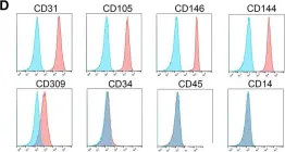
In Stem Cell Res Ther on 22 January 2019 by Gao, K., Kumar, P., et al.
Fig.1.D

-
FC/FACS
-
Homo sapiens (Human)
Collected and cropped from Stem Cell Res Ther by CiteAb, provided under a CC-BY license
Image 1 of 1