Severe traumatic injury has been associated with high susceptibility for the development of secondary complications caused by dysbalanced immune response. As the first line of the cellular immune response, neutrophils and monocytes recruited to the site of tissue damage and/or infection, are divided into three different subsets according to their CD16/CD62L and CD16/CD14 expression, respectively. Their differential functions have not yet been clearly understood. Thus, we evaluated the phenotypic changes of neutrophil and monocyte subsets among their functionality regarding oxidative burst and the phagocytic capacity in severely traumatized patients.
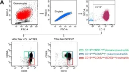
Peripheral blood was withdrawn from severely injured trauma patients (TP; n = 15, ISS ≥ 16) within the first 12 h post-trauma and from healthy volunteers (HV; n = 15) and stimulated with fMLP and PMA. CD16dimCD62Lbright (immature), CD16brightCD62Lbright (mature) and CD16brightCD62Ldim (CD62Llow) neutrophil subsets and CD14brightCD16- (classical), CD14brightCD16+ (intermediate) and CD14dimCD16+ (non-classical) monocyte subsets of HV and TP were either directly analyzed by flow cytometry or the examined subsets of HV were sorted first by fluorescence-activated cell sorting and subsequently analyzed. Subset-specific generation of reactive oxygen species (ROS) and of E. coli bioparticle phagocytosis were evaluated.
In TP, the counts of immature neutrophils were significantly increased vs. HV. The numbers of mature and CD62Ldim neutrophils remained unchanged but the production of ROS was significantly enhanced in TP vs. HV and the stimulation with fMLP significantly increased the generation of ROS in the mature and CD62Ldim neutrophils of HV. The counts of phagocyting neutrophils did not change but the mean phagocytic capacity showed an increasing trend in TP. In TP, the monocytes shifted toward the intermediate phenotype, whereas the classical and non-classical monocytes became less abundant. ROS generation was significantly increased in all monocyte subsets in TP vs. HV and PMA stimulation significantly increased those level in both, HV and TP. However, the PMA-induced mean ROS generation was significantly lower in intermediate monocytes of TP vs. HV. Sorting of monocyte and neutrophil subsets revealed a significant increase of ROS and decrease of phagocytic capacity vs. whole blood analysis.
Neutrophils and monocytes display a phenotypic shift following severe injury. The increased functional abnormalities of certain subsets may contribute to the dysbalanced immune response and attenuate the antimicrobial function and thus, may represent a potential therapeutic target. Further studies on isolated subsets are necessary for evaluation of their physiological role after severe traumatic injury.
Product Citations: 8
Severe Traumatic Injury Induces Phenotypic and Functional Changes of Neutrophils and Monocytes.
In Journal of Clinical Medicine on 14 September 2021 by Janicova, A., Becker, N., et al.
-
FC/FACS
In International Journal of Molecular Sciences on 26 July 2021 by Ahlmann, A. H., Fang, S., et al.
Small diameter (<6 mm) vessel grafts still pose a challenge for scientists worldwide. Decellularised umbilical artery (dUA) remains promising as small diameter tissue engineered vascular graft (TEVG), yet their immunogenicity remains unknown. Herein, we evaluated the host immune responses, with a focus on the innate part, towards human dUA implantation in mice, and confirmed our findings in an ex vivo allogeneic human setup. Overall, we did not observe any differences in the number of circulating white blood cells nor the number of monocytes among three groups of mice (1) dUA patch; (2) Sham; and (3) Mock throughout the study (day -7 to 28). Likewise, we found no difference in systemic inflammatory and anti-inflammatory cytokine levels between groups. However, a massive local remodelling response with M2 macrophages were observed in the dUA at day 28, whereas M1 macrophages were less frequent. Moreover, human monocytes from allogeneic individuals were differentiated into macrophages and exposed to lyophilised dUA to maximize an eventual M1 response. Yet, dUA did not elicit any immediate M1 response as determined by the absence of CCR7 and CXCL10. Together this suggests that human dUA elicits a minimal pro-inflammatory response further supporting its use as a TEVG in an allogeneic setup.
-
FC/FACS
-
Homo sapiens (Human)
-
Cardiovascular biology
-
Immunology and Microbiology
Eosinophil-derived neurotoxin: A biologically and analytically attractive asthma biomarker.
In PLoS ONE on 11 February 2021 by Rutten, B., Young, S., et al.
There is a growing body of evidence for the utility of eosinophil-derived neurotoxin (EDN) as a biomarker in asthma, including association with eosinophilic airway inflammation, assessment of disease severity and potential for predicting pathogenic risks, including exacerbations. However, to interpret any biomarker data with confidence, it is first important to understand the preanalytical factors and biological variation that may affect its reliable measurement and results interpretation. In this study we defined the healthy serum EDN reference range for men and women as 1.98 to 26.10 ng/mL, with no significant gender differences. Smoking did not impact the mean EDN levels and no circadian rhythm was identified for EDN, unlike blood eosinophils (EOS) where levels peaked at 00:00h. EDN expression in different cell types was investigated and shown to occur primarily in eosinophils, indicating they are likely to be the main cellular repository for EDN. We also confirm that the quantification of serum EDN is not influenced by the type of storage tube used, and it is stable at ambient temperature or when refrigerated for at least 7 days and for up to one year when frozen at -20°C or -80°C. In summary, EDN is a stable biomarker that may prove useful in precision medicine approaches by enabling the identification of a subpopulation of asthma patients with activated eosinophils and a more severe form of the disease.
-
Homo sapiens (Human)
Genetic landscape and autoimmunity of monocytes in developing Vogt-Koyanagi-Harada disease.
In Proceedings of the National Academy of Sciences of the United States of America on 13 October 2020 by Hu, Y., Hu, Y., et al.
Vogt-Koyanagi-Harada (VKH) disease is a systemic autoimmune disorder affecting multiple organs, including eyes, skin, and central nervous system. It is known that monocytes significantly contribute to the development of autoimmune disease. However, the subset heterogeneity with unique functions and signatures in human circulating monocytes and the identity of disease-specific monocytic populations remain largely unknown. Here, we employed an advanced single-cell RNA sequencing technology to systematically analyze 11,259 human circulating monocytes and genetically defined their subpopulations. We constructed a precise atlas of human blood monocytes, identified six subpopulations-including S100A12, HLA, CD16, proinflammatory, megakaryocyte-like, and NK-like monocyte subsets-and uncovered two previously unidentified subsets: HLA and megakaryocyte-like monocyte subsets. Relative to healthy individuals, cellular composition, gene expression signatures, and activation states were markedly alternated in VKH patients utilizing cell type-specific programs, especially the CD16 and proinflammatory monocyte subpopulations. Notably, we discovered a disease-relevant subgroup, proinflammatory monocytes, which showed a discriminative gene expression signature indicative of inflammation, antiviral activity, and pathologic activation, and converted into a pathologic activation state implicating the active inflammation during VKH disease. Additionally, we found the cell type-specific transcriptional signature of proinflammatory monocytes, ISG15, whose production might reflect the treatment response. Taken together, in this study, we present discoveries on accurate classification, molecular markers, and signaling pathways for VKH disease-associated monocytes. Therapeutically targeting this proinflammatory monocyte subpopulation would provide an attractive approach for treating VKH, as well as other autoimmune diseases.
-
FC/FACS
-
Homo sapiens (Human)
-
Genetics
-
Immunology and Microbiology
In International Journal of Molecular Sciences on 22 September 2018 by Tvedt, T. H. A., Melve, G. K., et al.
Interleukin-6 (IL-6) contributes to the development of immune-mediated complications after allogeneic stem cell transplantation. However, systemic IL-6 levels also increase during granulocyte colony-stimulating factor (G-CSF) mobilization of hematopoietic stem cells in healthy donors, but it is not known whether this mobilization alters systemic levels of other IL-6 family cytokines/receptors and whether such effects differ between donors. We examined how G-CSF administration influenced C-reactive protein (CRP) levels (85 donors) and serum levels of IL-6 family cytokines/receptors (20 donors). G-CSF increased CRP levels especially in elderly donors with high pretherapy levels, but these preharvesting levels did not influence clinical outcomes (nonrelapse mortality, graft versus host disease). The increased IL-6 levels during G-CSF therapy normalized within 24 h after treatment. G-CSF administration did not alter serum levels of other IL-6-familly mediators. Oncostatin M, but not IL-6, showed a significant correlation with CRP levels during G-CSF therapy. Clustering analysis of mediator levels during G-CSF administration identified two donor subsets mainly characterized by high oncostatin M and IL-6 levels, respectively. Finally, G-CSF could increase IL-6 release by in vitro cultured monocytes, fibroblasts, and mesenchymal stem cells. In summary, G-CSF seems to induce an acute phase reaction with increased systemic IL-6 levels in healthy stem cell donors.
-
Cardiovascular biology
-
Immunology and Microbiology
-
Stem Cells and Developmental Biology
In J Clin Med on 14 September 2021 by Janicova, A., Becker, N., et al.
Fig.1.A

-
FC/FACS
-
Collected and cropped from J Clin Med by CiteAb, provided under a CC-BY license
Image 1 of 1